. 1998 Oct 27;95(22):13153-8.
doi: 10.1073/pnas.95.22.13153.

HIV-1 Tat protein mimicry of chemokines

Affiliations

HIV-1 Tat protein mimicry of chemokines

A Albini et al. Proc Natl Acad Sci U S A. .

Abstract

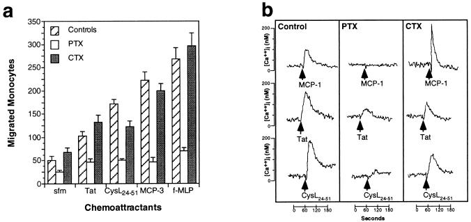
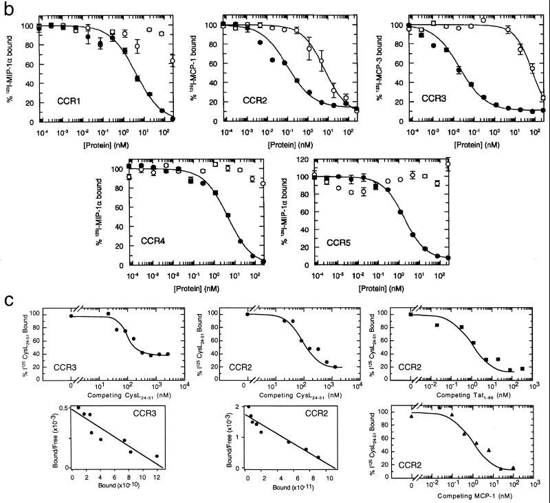
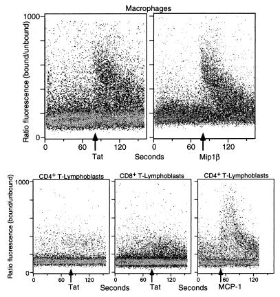
The HIV-1 Tat protein is a potent chemoattractant for monocytes. We observed that Tat shows conserved amino acids corresponding to critical sequences of the chemokines, a family of molecules known for their potent ability to attract monocytes. Synthetic Tat and a peptide (CysL24-51) encompassing the "chemokine-like" region of Tat induced a rapid and transient Ca2+ influx in monocytes and macrophages, analogous to beta-chemokines. Both monocyte migration and Ca2+ mobilization were pertussis toxin sensitive and cholera toxin insensitive. Cross-desensitization studies indicated that Tat shares receptors with MCP-1, MCP-3, and eotaxin. Tat was able to displace binding of beta-chemokines from the beta-chemokine receptors CCR2 and CCR3, but not CCR1, CCR4, and CCR5. Direct receptor binding experiments with the CysL24-51 peptide confirmed binding to cells transfected with CCR2 and CCR3. HIV-1 Tat appears to mimic beta-chemokine features, which may serve to locally recruit chemokine receptor-expressing monocytes/macrophages toward HIV producing cells and facilitate activation and infection.


Figures

Figure 1
Figure 1
Alignment of the HIV-1 Tat protein with the mature peptide sequences of the β-chemokines MCP-3, MCP-2, MCP-1, RANTES, MIP-1α, and MIP-1β. Residues conserved between Tat and the β-chemokines shown are shaded. The location of the Tat peptides along the Tat sequence used are indicated by brackets. The residues shown in italics (amino acids 87–101), not present in some HIV isolates, were not included in the Tat1–86 synthetic peptide used here.
Figure 2
Figure 2
(a) Induction of Ca2+ mobilization in monocytes by increasing doses of Tat1–86. As little as 100 ng⋅ml−1 (6.6 nM) produced a significant Ca2+ mobilization. (b) Inhibition of Tat1–86 induced Ca2+ mobilization by anti-Tat monoclonal antibodies (Anti Tat Ab) or trypsin digestion of Tat1–86. (c) Tat1–86 induced Ca2+ mobilization in different cell types. Tat (20 nM) induced rapid and transient Ca2+ mobilization in monocytes and in the monocyte-related THP-1 cell line, but not in T-lymphoblasts. Responses of these cells in the same experiment to a known chemokine (10 nM MCP-3) are shown for comparison. (d) Effect of peptides encompassing different domains of Tat on Ca2+ mobilization in monocytes. The cysteine-rich and core domain peptide (CysL24–51) produced a lower calcium mobilization as compared with the same molar concentration of Tat1–86 (100 nM). Higher concentrations of this peptide did produce a strong response. A peptide corresponding to the cysteine-rich domain (Cys20–39, 100 nM) showed a weaker, but still significant, activity. A peptide corresponding to CysL24–51, but with a mutation of the CCF sequence to SSG (CysL24–51CCFmut) showed no activity. Peptides corresponding to the RGD and Basic domains were also ineffective at the doses shown or even 20-fold higher doses (2 μM, not shown). The effects of RANTES is shown for comparison.
Figure 3
Figure 3
(a) Effects of pertussis toxin (PTX) and cholera toxin (CTX) on monocyte migration in response to 400 ng ml−1 Tat1–86, 1 μg⋅ml−1 CysL24–51, and 100 ng⋅ml−1 MCP-3 and f-MLP. PTX caused the marked inhibition of chemotaxis to all the chemoattractants tested, CTX did not substantially affect migration. (b) Effects of PTX and CTX on calcium mobilization induced by 10 nM MCP-1, 20 nM Tat1–86, or 100 nM CysL24–51 peptide in monocytes.
Figure 4
Figure 4
(a) Cross-desensitization experiments with Tat1–86, the CysL24–51 peptide, and some β-chemokines on monocytes. The response to the ligand (indicated at the top of each panel) is given as the percent of maximal response to the same concentration of ligand without desensitization. The concentrations of the desensitization agent are indicated on the abscissa. −, indicates no desensitization agent added. (b) Equilibrium competition on cell membranes from Chinese hamster ovary cells expressing CCR1, CCR2, CCR3, or CCR5, and HEK 293 cells expressing CCR4, as indicated. The competition by Tat1–86 (○) or unlabeled chemokine (•) was determined by a scintillation proximity assay binding assay as described (22). The radiolabeled and unlabeled chemokines were MIP-1α for CCR1, CCR4, and CCR5; MCP-1 for CCR2; and MCP-3 for CCR3. (c) Competitive ligand binding of radiolabeled CysL24–51 peptide to transfected cells expressing CCR2 or CCR3, as indicated. The competition for 125I-CysL24–51 peptide binding by unlabeled CysL24–51 peptide (•), Tat (■), or MCP-1 (▴) was determined. Scatchard plot of unlabeled CysL24–51 peptide competition was calculated from the data and presented below binding curves.
Figure 4
Figure 4
(a) Cross-desensitization experiments with Tat1–86, the CysL24–51 peptide, and some β-chemokines on monocytes. The response to the ligand (indicated at the top of each panel) is given as the percent of maximal response to the same concentration of ligand without desensitization. The concentrations of the desensitization agent are indicated on the abscissa. −, indicates no desensitization agent added. (b) Equilibrium competition on cell membranes from Chinese hamster ovary cells expressing CCR1, CCR2, CCR3, or CCR5, and HEK 293 cells expressing CCR4, as indicated. The competition by Tat1–86 (○) or unlabeled chemokine (•) was determined by a scintillation proximity assay binding assay as described (22). The radiolabeled and unlabeled chemokines were MIP-1α for CCR1, CCR4, and CCR5; MCP-1 for CCR2; and MCP-3 for CCR3. (c) Competitive ligand binding of radiolabeled CysL24–51 peptide to transfected cells expressing CCR2 or CCR3, as indicated. The competition for 125I-CysL24–51 peptide binding by unlabeled CysL24–51 peptide (•), Tat (■), or MCP-1 (▴) was determined. Scatchard plot of unlabeled CysL24–51 peptide competition was calculated from the data and presented below binding curves.
Figure 5
Figure 5
Ca2+ fluxes generated by Tat on monocyte-derived macrophages or CD4+ or CD8+ T-lymphoblasts (as indicated) assessed by multiparameter flow cytometry analysis. The response of monocytes to MIP-1β and of CD4+ T-lymphoblasts to MCP-1 is shown for comparison.

References

    1. Brake D, Debouck C, Biesecker G. J Cell Biol. 1990;111:1275–1281. - PMC - PubMed
    1. Albini A, Benelli R, Presta M, Rusnati M, Ziche M, Rubartelli A, Paglialunga G, Bussolino F, Noonan D. Oncogene. 1996;12:289–297. - PubMed
    1. Lafrenie R M, Wahl L M, Epstein J S, Hewlett I K, Yamada K M, Dhawan S. J Immunol. 1996;157:974–977. - PubMed
    1. Mitola S, Sozzani S, Luini W, Primo L, Bosatti A, Weich H, Bussolino F. Blood. 1997;90:1365–1372. - PubMed
    1. Benelli R, Mortarini R, Anichini A, Giunciuglio D, Noonan D M, Montalti S, Tacchetti C, Albini A. AIDS. 1998;12:261–268. - PubMed

Publication types

MeSH terms