. 1998 Oct 27;95(22):13182-7.
doi: 10.1073/pnas.95.22.13182.

Inhibition of arachidonate 5-lipoxygenase triggers massive apoptosis in human prostate cancer cells

Affiliations

Inhibition of arachidonate 5-lipoxygenase triggers massive apoptosis in human prostate cancer cells

J Ghosh et al. Proc Natl Acad Sci U S A. .

Abstract

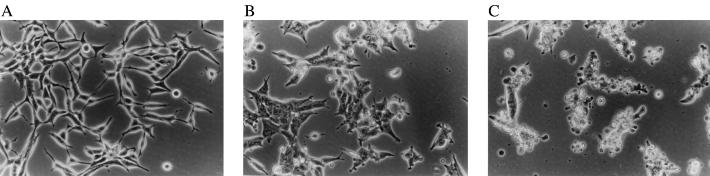
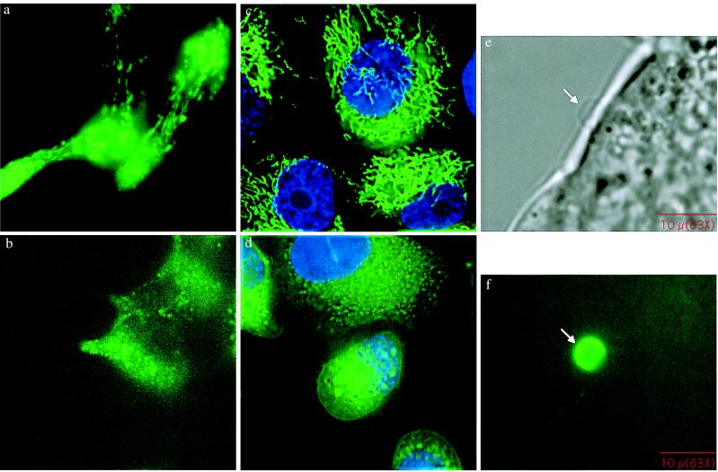
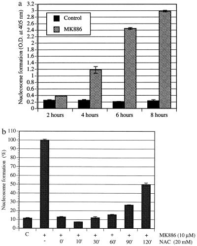
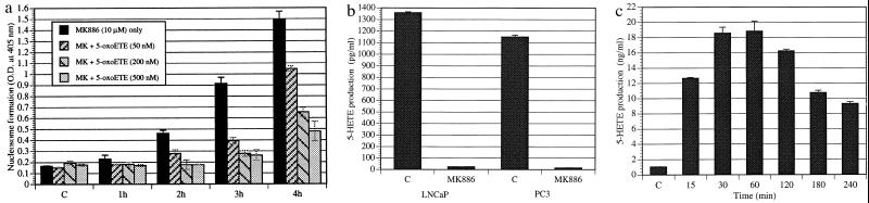
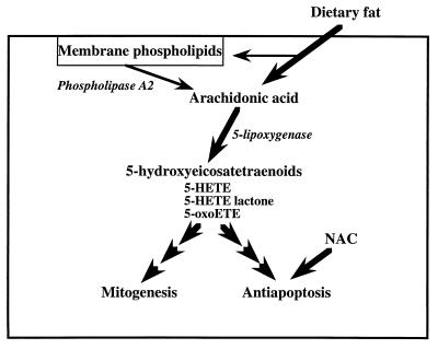
Diets high in fat are associated with an increased risk of prostate cancer, although the molecular mechanism is still unknown. We have previously reported that arachidonic acid, an omega-6 fatty acid common in the Western diet, stimulates proliferation of prostate cancer cells through production of the 5-lipoxygenase metabolite, 5-HETE (5-hydroxyeicosatetraenoic acid). We now show that 5-HETE is also a potent survival factor for human prostate cancer cells. These cells constitutively produce 5-HETE in serum-free medium with no added stimulus. Exogenous arachidonate markedly increases the production of 5-HETE. Inhibition of 5-lipoxygenase by MK886 completely blocks 5-HETE production and induces massive apoptosis in both hormone-responsive (LNCaP) and -nonresponsive (PC3) human prostate cancer cells. This cell death is very rapid: cells treated with MK886 showed mitochondrial permeability transition between 30 and 60 min, externalization of phosphatidylserine within 2 hr, and degradation of DNA to nucleosomal subunits beginning within 2-4 hr posttreatment. Cell death was effectively blocked by the thiol antioxidant, N-acetyl-L-cysteine, but not by androgen, a powerful survival factor for prostate cancer cells. Apoptosis was specific for 5-lipoxygenase-programmed cell death was not observed with inhibitors of 12-lipoxygenase, cyclooxygenase, or cytochrome P450 pathways of arachidonic acid metabolism. Exogenous 5-HETE protects these cells from apoptosis induced by 5-lipoxygenase inhibitors, confirming a critical role of 5-lipoxygenase activity in the survival of these cells. These findings provide a possible molecular mechanism by which dietary fat may influence the progression of prostate cancer.


Figures

Figure 1
Figure 1
Photomicrographs showing the morphology of LNCaP prostate cancer cells. LNCaP prostate cancer cells (3 × 10⁵) were grown for 48 hr in 60-mm tissue culture plates in RPMI medium 1640 supplemented with 10% FBS. The spent medium was then replaced with fresh serum-free RPMI medium 1640, and the cells were treated with 10 μM MK886 for various periods of time. Control cells were treated with the untreated medium containing 0.02% DMSO. Photographs were taken with a Zeiss inverted microscope at ×20. (A) Control cells. (B and C) Cells 4 and 8 hr, respectively, after treatment with MK886.
Figure 2
Figure 2
(ad) Fluorescent microscope images showing MPT. LNCaP and PC3 cells (3 × 10⁵ per well) were plated on cover glasses in 6-well tissue culture plates in RPMI medium 1640 supplemented with 10% FBS and cultured for 48 hr. The cover glasses were then placed in 6-well plates in 1 ml of RPMI medium 1640 and loaded with 5 μM Rhodamine-123 for 30 min. The cells were then treated with 10 μM MK886, incubated for 45 min at 37°C, and observed by fluorescent microscopy. (a and b) LNCaP cells, (c and d) PC3 cells. (a and c) Control. (b) and (d) MK886-treated. (e) and (f) Fluorescent microscope images showing phosphatidylserine externalization. LNCaP prostate cancer cells were plated as described and treated with 10 μM MK886 in serum-free medium for 3 hr. At the end of each incubation period, cells were washed and treated with annexin V-fluorescein isothiocyanate in binding buffer for 15 min at room temperature. The cover glasses were then mounted on glass slides and observed by fluorescent microscopy using fluorescein long pass filter set. e and f represent images of a bleb under phase-contrast and fluorescent microscope, respectively.
Figure 3
Figure 3
(a) Time course of the formation of nucleosomal DNA in prostate cancer cells on treatment with MK886. LNCaP prostate cancer cells (3 × 10⁵) were cultured for 48 hr in 60-mm tissue culture plates in RPMI medium 1640 supplemented with 10% FBS. The medium was then changed to serum-free RPMI medium 1640, and the cells were treated with 10 μM MK886 for various periods of time. Control cells were treated with the untreated medium containing 0.02% DMSO only. At the end of each incubation periods, cells were harvested and lysed, and the formation of nucleosomes was quantitatively measured by Cell Death Detection ELISA taking lysates equivalent to 5,000 cells for each assay. Results are presented as the mean ± standard error (n = 4). (b) Effect of NAC on MK886-induced prostate cancer cell death. LNCaP prostate cancer cells were plated as described in Fig. 3a and treated with 10 μM MK886 in serum-free medium. The thiol antioxidant NAC (20 mM), was added at different time points as indicated (minutes) post-MK886 treatment. Control cells were treated with the plating medium containing the solvent (0.02% DMSO) only. Cells were incubated for 6 hr after the addition of MK886, and the formation of nucleosomes was measured by Cell Death Detection ELISA. Data presented as the mean ± standard error (n = 4).
Figure 4
Figure 4
(a) Reversal of MK886-induced DNA degradation by 5-oxoETE. Cells in serum-free medium were treated with 10 μM MK886 as described in Fig. 3a in the absence or presence of the 5-lipoxygenase product, 5-oxoETE, and incubated for various periods of time at 37°C. Control cells (C) were treated with serum-free RPMI medium 1640 containing 0.02% DMSO. Formation of nucleosomal DNA was determined by Cell Death Detection ELISA. Results represent the mean ± standard error (n = 4). (b) Constitutive production of 5-HETE by human prostate cancer cells. Cells (106) were plated in serum-free RPMI medium 1640 in 60-mm tissue culture dishes and incubated for 24 hr. Then the cells were treated with 10 μM MK886 or the solvent vehicle (0.02% DMSO) and incubated at 37°C for 2 hr. At the end of the incubation period, aliquots of culture supernatants were taken, and the production of 5-HETE was measured by radioimmunoassay. Each data point represents the mean ± standard error (n = 4). (c) Stimulation of 5-HETE production in LNCaP cells by arachidonic acid. Cells were plated as described in b and treated with 10 μM arachidonic acid precomplexed with lipid-free BSA for various periods of time. Production of 5-HETE was measured by radioimmunoassay using aliquots of culture supernatant. Data is presented as the mean ± standard error (n = 4).
Figure 5
Figure 5
A model illustrating regulation by dietary fat of prostate cancer cell growth and survival.

References

    1. Parker S L, Tong T, Bolden S, Wingo P A. Ca Cancer J Clin. 1997;47:5–27. - PubMed
    1. Crawford E D, Eisenberger M A, McLeod D G, Spaulding J T, Benson R, Dorr A, Blumenstein B A, Davis M A, Goodman P J. N Engl J Med. 1989;321:419–424. - PubMed
    1. Gann P H, Hennekens C H, Sacks F M, Grodstein F, Giovannucci E L, Stampfer M J. J Natl Cancer Inst. 1994;86:281–286. - PubMed
    1. West D W, Slattery M L, Robison L M, French T K, Mahoney A W. Cancer Causes Control. 1991;2:85–94. - PubMed
    1. Giovannucci E, Rimm E B, Colditz G A, Stampfer M J, Ascherio A, Chute C C, Willett W C. J Natl Cancer Inst. 1993;85:1571–1579. - PubMed

Publication types

MeSH terms