. 1998 Oct 27;95(22):13272-7.
doi: 10.1073/pnas.95.22.13272.

Ca2+/calmodulin-dependent kinase II mediates simultaneous enhancement of gap-junctional conductance and glutamatergic transmission

Affiliations

Ca2+/calmodulin-dependent kinase II mediates simultaneous enhancement of gap-junctional conductance and glutamatergic transmission

A E Pereda et al. Proc Natl Acad Sci U S A. .

Abstract

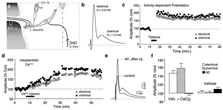
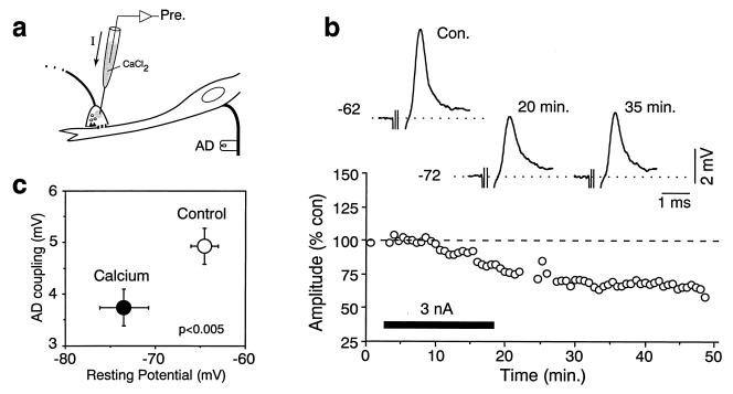
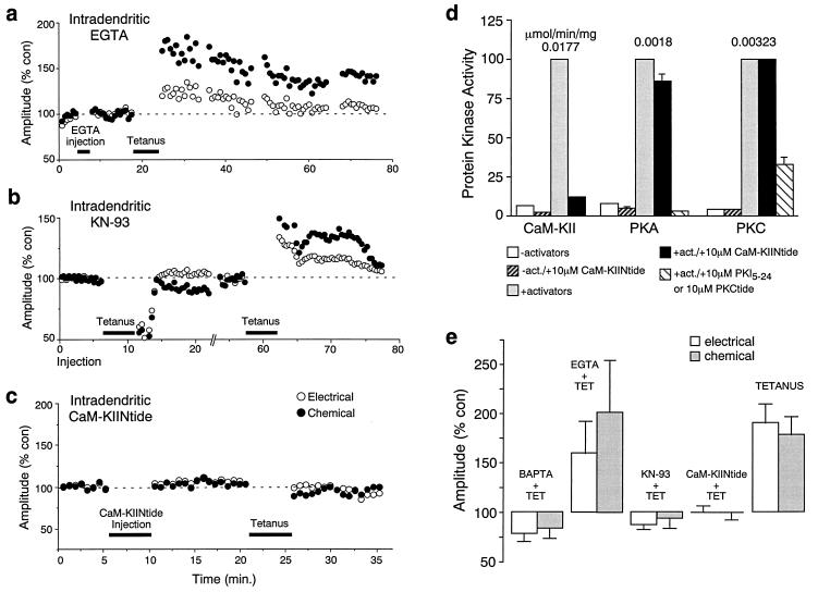
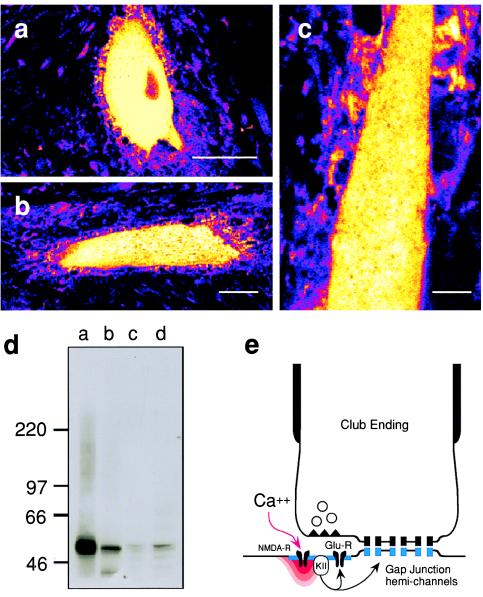
While chemical synapses are very plastic and modifiable by defined activity patterns, gap junctions, which mediate electrical transmission, have been classically perceived as passive intercellular channels. Excitatory transmission between auditory afferents and the goldfish Mauthner cell is mediated by coexisting gap junctions and glutamatergic synapses. Although an increased intracellular Ca2+ concentration is expected to reduce gap junctional conductance, both components of the synaptic response were instead enhanced by postsynaptic increases in Ca2+ concentration, produced by patterned synaptic activity or intradendritic Ca2+ injections. The synaptically induced potentiations were blocked by intradendritic injection of KN-93, a Ca2+/calmodulin-dependent kinase (CaM-K) inhibitor, or CaM-KIINtide, a potent and specific peptide inhibitor of CaM-KII, whereas the responses were potentiated by injection of an activated form of CaM-KII. The striking similarities of the mechanisms reported here with those proposed for long-term potentiation of mammalian glutamatergic synapses suggest that gap junctions are also similarly regulated and indicate a primary role for CaM-KII in shaping and regulating interneuronal communication, regardless of its modality.


Figures

Figure 1
Figure 1
Mixed synapses on the Mauthner cell (M-cell) exhibit activity-dependent potentiations. (a) Experimental arrangement (see Material and Methods). V, voltage; P, pressure; 8th n., eighth nerve; L. dendrite, lateral dendrite; AD, antidromic stimulation of the spinal cord (S. cord). (b) Eighth nerve stimulation evokes a fast electrotonic potential followed by a chemical glutamatergic excitatory postsynaptic potential (EPSP), with the indicated decay time constants. (c) Discontinuous tetanic stimulation of the nerve produces persistent homosynaptic potentiations of both components. Plots here and in subsequent figures illustrate the amplitudes of the electrotonic (○) and chemical ([circf) components versus time (each point represents the average of 20 traces) for one experiment. (d) Intradendritic injections of Ca2+ enhanced both components. (e) Superimposed traces represent the averages of 20 consecutive responses obtained in the control and 40 min after Ca2+ injection, at the maximum level of potentiation. (f) Bar plots represent the amplitudes (% of control) of the electrotonic potential, chemical EPSP, and antidromic spike (AD), all measured once the maximum potentiation had been reached. They averaged 146.4% (±7.2%) and 158.6% (±11.8%) of control for the electrical and chemical components, respectively (n = 5). Antidromic spike height, a measure of the cell’s input resistance, remained unchanged (98.8% ± 3.8%). Recordings made with the vehicle solution in the electrode did not affect the amplitudes, as measured at comparable time intervals, averaging 102.9% ± 2.8%, 101.6% ± 7%, and 109.4% ± 4.6%, for the electrical and chemical postsynaptic potentials and the AD spike, respectively (n = 5). Here and elsewhere, error bars represent 1 SEM.
Figure 2
Figure 2
Presynaptic injections of Ca2+ do not increase junctional conductance. (a) Intraterminal recordings were obtained from large myelinated club endings, and Ca2+ was injected iontophoretically. (b) After Ca2+ injection the terminal was hyperpolarized from −62 to −72 mV in this case, and the antidromic coupling potential was decreased to approximately 70% of control (Upper). (Lower) Plot of the amplitude of the antidromic coupling potential versus time (each point represents the average of 10 traces) for the same experiment. (c) Diagram summarizing the values of the resting potential and coupling potential amplitudes (AD coupling) obtained for 11 terminals in control (○) and after at least 10 min of continuous Ca2+ injection (•). The changes were statistically significant.
Figure 3
Figure 3
Evidence that Ca2+ effects are mediated by CaM-KII. (a) Intradendritic EGTA injections did not prevent the induction of potentiations by tetanic eighth nerve stimulation. (b) Intradendritic injection of KN-93 blocked induction. In this experiment KN-93 was pressure injected for 3 min, ending at time 0, and PSP amplitudes postinjection were taken as controls. The antagonist itself had no significant effect. While tetanic stimulation 7–11 min after the injection failed (typically a transient depression followed unsuccessful tetani), a second tetanus 1 hr later successfully triggered potentiations of both components (each point represents the average of 15 traces). A new control was established at 50 min. (c) Intradendritic injections of CaM-KIINtide prevented the induction. (d) Specificity of CaM-KIINtide for CaM-KII in goldfish brain: Bar plots of protein kinase activity in the indicated conditions. CaM-KII was assayed for 1 min, PKA and PKC for 8 min each. Each column represents a mean and standard deviation of three data points. (e) To accurately estimate the effects of intradendritically injected compounds on the induction of these rapid activity-dependent potentiations, averages of the last 15–40 responses obtained before and after tetanic stimulation were compared. Bar plots represent the normalized posttetanus amplitude (% of control) of the electrotonic potential and chemical EPSP. BAPTA and EGTA at 5 mM have significantly different (P < 0.05) effects on induction (BAPTA + TET, EGTA + TET). In the presence of BAPTA, tetanic stimulation transiently depressed both PSPs, which averaged 78.1% (±9.8%, SEM) for the electrical and 83.8% (±7.4%, SEM) for the chemical, of their respective control amplitudes (n = 5; ref. 4). In the presence of EGTA the two components averaged 159.6% (±33.1%) and 202.6% (±53.2%), of their respective control amplitudes (n = 5). Intradendritic injections of KN-93 blocked activity-dependent potentiations (KN-93 + TET). Electrical and chemical responses averaged 86.8% (±4.4%) and 93.7% (±10.5%), of their respective control amplitudes (n = 9). CaM-KIINtide also prevented induction of the potentiations (CaM-KIINtide + TET; n = 12). In contrast, tetanic stimulation in controls (n = 30) produced potentiations that averaged 190.02% (±19.4%) and 178.8% (±18.2%).
Figure 4
Figure 4
Intradendritic injections of a constitutively active form of α-CaM-KII enhanced both components of the synaptic response. (a) Time course of two experiments in which recordings were obtained with electrodes containing either the constitutively active or a heat-inactivated form of α-CaM-KII. (b) Superimposed average traces (>20) obtained at different intervals after penetrating the dendrite with an electrode containing α-CaM-KII. Typically, leakage of α-CaM-KII alone caused continuously increasing potentiations. (c) Bar plots represent the normalized amplitudes of the synaptic components and antidromic (AD) spike measured at the end of the recording sessions in experiments with α-CaM-KII (30–80 min; electrical and chemical PSPs averaged 152.1% ± 6.5% and 158.3% ± 16.7% of their respective control amplitudes, respectively, n = 7) and heat-inactivated α-CaM-KII (40–80 min; corresponding synaptic responses averaged 98.3% ± 6.6% and 96% ± 7.4%, n = 7). M-cell antidromic spike height did not change significantly in either experimental series.
Figure 5
Figure 5
CaM-KII immunoreactivity is present in goldfish M-cells. (a–c) Immunohistochemical evidence for the presence of CaM-KII in the M-cell. Confocal (pseudocolor; white corresponds to maximum brightness) images obtained with the antibody G-301 (1:1000; Texas red). (a) Soma. (Bar: 50 μm.) (b) Lateral dendrite. (Bar: 30 μm.) (c) Higher magnification view of another stained lateral dendrite. (Bar: 15 μm.) Note the punctate staining surrounding the M-cell soma and lateral dendrite, most likely corresponding to the presynaptic localization of this enzyme. (d) Immunoblot using G-301 (1:200 dilution) and the following: lane a, 2 μg of rat cerebral cortex homogenate; lane b, 2.5 ng of CaM-KII purified from rat forebrain (11); lanes c and d, 30 μg and 90 μg, respectively, of goldfish brain homogenate. Molecular weight markers (× 10−3) are shown on the left. (e) Schematic representation of the proposed potentiating pathway. KII, CaM-KII; R, receptor.

References

    1. Malenka R C, Nicoll R A. Trends Neurosci. 1993;16:521–527. - PubMed
    1. Bennett M V L. In: Cellular Biology of Neurons, Handbook of Physiology. Kandel E R, editor. 1, Sect. I. Baltimore: Williams and Wilkins; 1977. pp. 357–416.
    1. Yang X D, Korn H, Faber D S. Nature (London) 1990;348:542–545. - PubMed
    1. Pereda A, Faber D S. J Neurosci. 1996;16:983–992. - PMC - PubMed
    1. Bartelmez G W. J Comp Neurol. 1915;25:87–128.

Publication types

MeSH terms

LinkOut - more resources