. 1998 Oct 27;95(22):13278-83.
doi: 10.1073/pnas.95.22.13278.

A Rap guanine nucleotide exchange factor enriched highly in the basal ganglia

Affiliations

A Rap guanine nucleotide exchange factor enriched highly in the basal ganglia

H Kawasaki et al. Proc Natl Acad Sci U S A. .

Erratum in

  • Proc Natl Acad Sci U S A 1999 Jan 5;96(1):318

Abstract

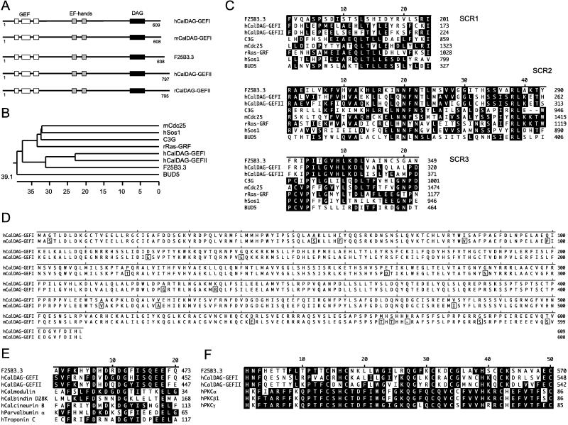
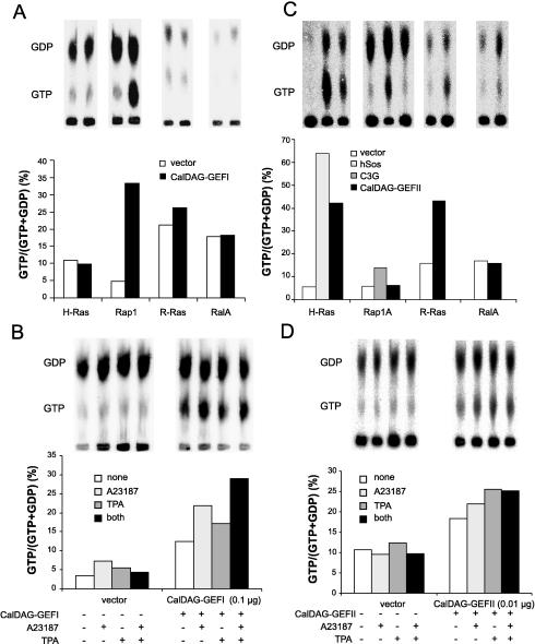
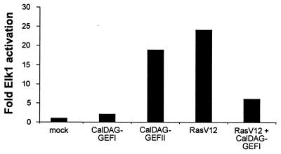
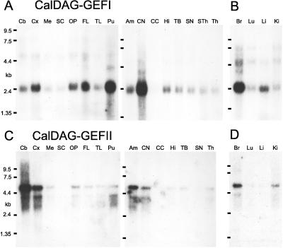
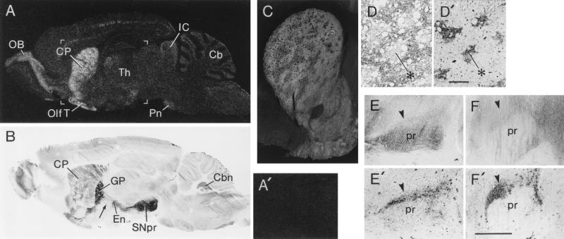
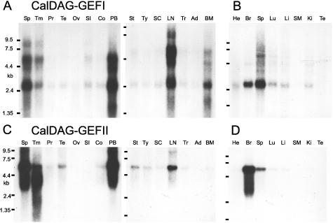
Ras proteins, key regulators of growth, differentiation, and malignant transformation, recently have been implicated in synaptic function and region-specific learning and memory functions in the brain. Rap proteins, members of the Ras small G protein superfamily, can inhibit Ras signaling through the Ras/Raf-1/mitogen-activated protein (MAP) kinase pathway or, through B-Raf, can activate MAP kinase. Rap and Ras proteins both can be activated through guanine nucleotide exchange factors (GEFs). Many Ras GEFs, but to date only one Rap GEF, have been identified. We now report the cloning of a brain-enriched gene, CalDAG-GEFI, which has substrate specificity for Rap1A, dual binding domains for calcium (Ca2+) and diacylglycerol (DAG), and enriched expression in brain basal ganglia pathways and their axon-terminal regions. Expression of CalDAG-GEFI activates Rap1A and inhibits Ras-dependent activation of the Erk/MAP kinase cascade in 293T cells. Ca2+ ionophore and phorbol ester strongly and additively enhance this Rap1A activation. By contrast, CalDAG-GEFII, a second CalDAG-GEF family member that we cloned and found identical to RasGRP [Ebinu, J. O., Bottorff, D. A., Chan, E. Y. W., Stang, S. L., Dunn, R. J. & Stone, J. C. (1998) Science 280, 1082-1088], exhibits a different brain expression pattern and fails to activate Rap1A, but activates H-Ras, R-Ras, and the Erk/MAP kinase cascade under Ca2+ and DAG modulation. We propose that CalDAG-GEF proteins have a critical neuronal function in determining the relative activation of Ras and Rap1 signaling induced by Ca2+ and DAG mobilization. The expression of CalDAG-GEFI and CalDAG-GEFII in hematopoietic organs suggests that such control may have broad significance in Ras/Rap regulation of normal and malignant states.


Figures

Figure 1
Figure 1
(A) CalDAG-GEFs represent a family of Ras-superfamily GEFs including human (h) and mouse (m) CalDAG-GEFI, human (h) and rat (r) CalDAG-GEFII, and Caenorhabditis elegans (F25B3.3, GenBank accession no. 1262950) CalDAG-GEF. (B) Computer-generated phylogenetic tree analysis of the GEF domains of CalDAG-GEFI and CalDAG-GEFII in relation to other Ras-superfamily GEFs. (C) Multiple alignment of GEF structurally conserved regions (SCRs) of CalDAG-GEFs and several other GEFs of the Ras superfamily. (D) Full-length amino acid sequence of human (h) and mouse (m) CalDAG-GEFI (box indicates amino acid differences). (E) Sequence similarity (black indicates identity) of EF-hand domains in CalDAG-GEFs and other calcium-binding proteins. (F) Sequence similarity of DAG-binding domains of CalDAG-GEFs and PKC (protein kinase C) family proteins. Multiple sequence alignments and phylogenetic tree analysis were carried out with the lasergene Software Package (DNAstar, Madison, WI). Abbreviations and GenBank accession nos. of protein sequences are: C3G, 474982; mCdc25, 882120; rRas-GRF, 57665; hSos1 (human son-of-sevenless 1), 476780; BUD5, 171141; hCalmodulin, 115512; hCalbindin D28k, 227666; hCalcineurin B, 105504; hParvalbumin α, 131100; hTroponin C, 136043; hPKCα, 125549; hPKCβ1, 125538; hPKCγ, 462455.
Figure 2
Figure 2
Contrasting functional properties of CalDAG-GEFI and CalDAG-GEFII. (A) Activation of Rap1 by CalDAG-GEFI in 293T cells expressing Ras-family small G proteins with or without CalDAG-GEFI. (B) Calcium ionophore and TPA enhancement of Rap1A activation by CalDAG-GEFI in 293T cells. (C) CalDAG-GEFII activates H-Ras and R-Ras in intact 293T cells. GST-tagged H-Ras, Rap1A, R-Ras, or RalA were expressed in 293T cells with or without Sos1, C3G, or CalDAG-GEFII. (D) Augmentation of H-Ras activation by CalDAG-GEFII in the presence of A23187 or TPA or both, assayed as in B.
Figure 3
Figure 3
Inhibition of Ras/Erk/Elk1 signaling cascade by CalDAG-GEFI and activation of this cascade by CalDAG-GEFII. 293T cells were transfected with pFR-Luc and pFA-Elk1 together with expression vectors as indicated.
Figure 4
Figure 4
Northern hybridization analysis of CalDAG-GEFI and CalDAG-GEFII in adult human brain. (A) Enriched expression of CalDAG-GEFI in the putamen and caudate nucleus. Compare with C, showing that human brain CalDAG-GEFII mRNA expression levels are most enriched in cerebellum, cerebral cortex, and amygdala. B and D show high brain expression in human fetus. Am, amygdala; Br, brain; Cb, cerebellum; CC, corpus callosum; CN, caudate nucleus; Cx, cortex; FL, frontal lobe; Hi, hippocampus; Ki, kidney; Li, liver; Lu, lung; Me, medulla oblongata; OP, occipital pole; Pu, putamen; SC, spinal cord; SN, substantia nigra; Sth, subthalamic nucleus; TB, total brain; Th, thalamus; TL, temporal lobe.
Figure 5
Figure 5
Preferential distribution of CalDAG-GEFI in the basal ganglia. (A) In situ hybridization film autoradiogram of parasagittal section through rat brain demonstrating predominant localization of CalDAG-GEFI mRNA in the striatum and olfactory bulb with weaker signal in other brain sites. (A′) Sense hybridization in control section shows no label in striatum. Area shown roughly corresponds to bracketed region in A. (B) Immunohistochemical staining of parasagittal section through rat brain with CalDAG-GEFI mAb 18B11 shows strong staining of striatum and striatal projection fibers, and intense staining of globus pallidus and substantia nigra, with significant staining also in olfactory bulb and brainstem (including cerebellar) nuclei. Arrow points to immunostained striatal projection fibers. (C) Reverse-contrast photomicrograph of CalDAG-GEFI immunoreactivity (which appears light) in rat striatum, demonstrating predominant expression of CalDAG-GEFI in the striatal matrix compartment. (D and D′) Correspondence of CalDAG-GEFI-poor zones (D) in rat caudoputamen with μ-opioid rich striosomes (D′, example at asterisks) stained in adjoining sections. (Bar = 200 μm.) (E–F′) Knife-cut through subthalamic region sharply decreases CalDAG-GEFI immunostaining in substantia nigra. Anterior to left. Arrowheads point to substantia nigra pars compacta. (E) Control side (with thalamic knife-cut sparing striatal projection fibers) shows strong CalDAG-GEFI immunostaining. (E′) Serially adjoining section stained for TH showing location of TH-positive substantia nigra pars compacta. (F) Reduced CalDAG-GEFI immunostaining in substantia nigra on side of subthalamic knife-cut. (F′) Serially adjoining section showing TH-positive substantia nigra pars compacta. (Bar = 1 mm.) Cb, cerebellum; Cbn, cerebellar nuclei; CP, caudoputamen; En, entopeduncular nucleus; GP, globus pallidus; IC, inferior colliculus; OB, olfactory bulb; Olf T, olfactory tubercle; Pn, pontine nuclei; pr, substantia nigra pars reticulata; SNpr, substantia nigra pars reticulata; Th, thalamus.
Figure 6
Figure 6
CalDAG-GEFI and CalDAG-GEFII are expressed in the hematopoietic system. (A and B) CalDAG-GEFI expression in adult human (A) and rat (B) organs. (C and D) CalDAG-GEFII expression in adult human (C) and rat (D) organs. Ad, adrenal gland; BM, bone marrow; Br, brain; Co, colon (mucosal lining); He, heart; Ki, kidney; Li, liver; LN, lymph node; Lu, lung; Ov, ovary; PB, peripheral blood leukocytes; Pr, prostate; SC, spinal cord; SI, small intestine; SM, skeletal muscle; Sp, spleen; St, stomach; Te, testis; Tm, thymus; Tr, trachea; Ty, thyroid.

References

    1. Graybiel A M. Neurobiol Learning Memory. 1998;70:119–136. - PubMed
    1. Knowlton B J, Mangels J A, Squire L R. Science. 1996;273:1399–1402. - PubMed
    1. Glatt C E, Snyder S H. Nature (London) 1993;361:536–538. - PubMed
    1. Drinnan S L, Hope B T, Snutch T P, Vincent S R. Mol Cell Neurosci. 1991;2:66–70. - PubMed
    1. Celio M R. Neuroscience. 1990;35:375–475. - PubMed

Publication types

MeSH terms