. 1998 Oct 27;95(22):13307-12.
doi: 10.1073/pnas.95.22.13307.

A G protein gamma subunit-like domain shared between RGS11 and other RGS proteins specifies binding to Gbeta5 subunits

Affiliations

A G protein gamma subunit-like domain shared between RGS11 and other RGS proteins specifies binding to Gbeta5 subunits

B E Snow et al. Proc Natl Acad Sci U S A. .

Abstract

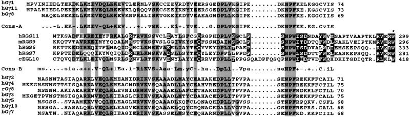
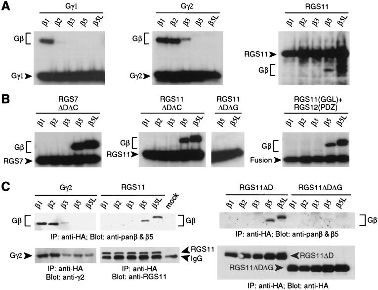
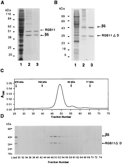
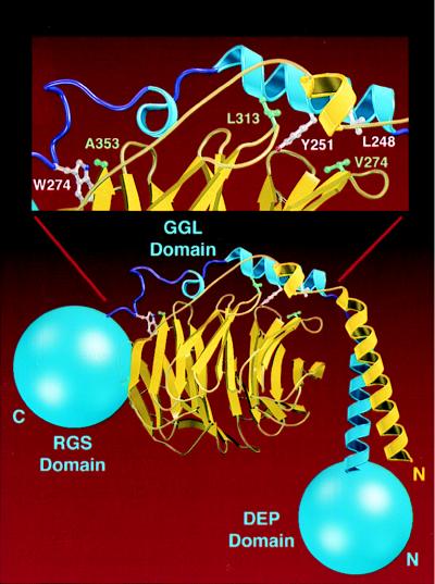
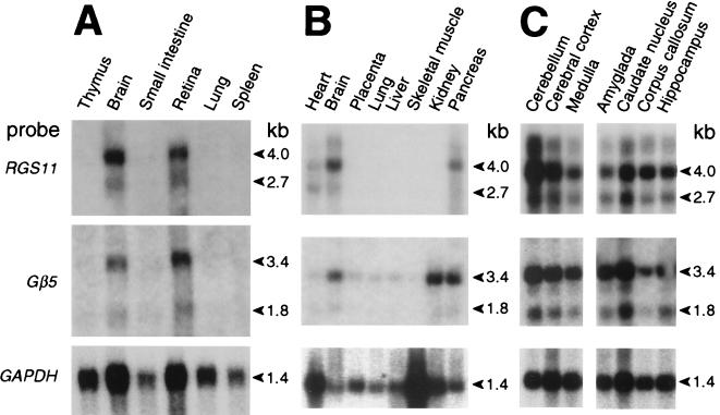
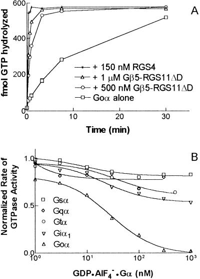
Regulators of G protein signaling (RGS) proteins act as GTPase-activating proteins (GAPs) toward the alpha subunits of heterotrimeric, signal-transducing G proteins. RGS11 contains a G protein gamma subunit-like (GGL) domain between its Dishevelled/Egl-10/Pleckstrin and RGS domains. GGL domains are also found in RGS6, RGS7, RGS9, and the Caenorhabditis elegans protein EGL-10. Coexpression of RGS11 with different Gbeta subunits reveals specific interaction between RGS11 and Gbeta5. The expression of mRNA for RGS11 and Gbeta5 in human tissues overlaps. The Gbeta5/RGS11 heterodimer acts as a GAP on Galphao, apparently selectively. RGS proteins that contain GGL domains appear to act as GAPs for Galpha proteins and form complexes with specific Gbeta subunits, adding to the combinatorial complexity of G protein-mediated signaling pathways.


Figures

Figure 1
Figure 1
Sequence alignment between Gγ subunits and the GGL domains of RGS6, RGS7, RGS9, RGS11, and EGL-10. Residues conserved among RGS proteins are in black boxes; residues in common between RGS proteins and one or both Gγ consensus lines (Cons-A, Cons-B) are shown in shaded boxes. Asterisk denotes first residue of the RGS domain. b, bovine; c, C. elegans; h, human; m, mouse; r, rat.
Figure 2
Figure 2
Gβ binding specificity of the GGL domain. HA-tagged Gγ or RGS proteins were either cotranslated in vitro (A and B) or cotransfected into COS-7 cells (C) with individual Gβ subunits, immunoprecipitated (IP) with anti-HA mAb, and visualized by SDS/PAGE and autoradiography (A and B) or immunoblotting (C) with indicated antisera (Blot). (A) Gβ subunit association in vitro with Gγ1, Gγ2, and full-length RGS11 proteins. (B) Gβ subunit association in vitro with truncated RGS7 protein (ΔDΔC, aa 202–395 of SwissProt accession no.P49802), truncated RGS11 proteins (ΔDΔC, aa 219–423; ΔDΔG, aa 283–467), and a chimeric protein (Fusion) composed of the RGS11 GGL domain (aa 219–283) fused to the rat RGS12 PDZ domain (aa 1–91 of SwissProt accession no. O08774). (C) Gβ subunit association with Gγ2, full-length RGS11, and truncated RGS11 proteins (ΔD, aa 219–467; ΔDΔG, aa 283–467) in COS-7 cells.
Figure 3
Figure 3
Purification of Gβ5/RGS11 heterodimers after expression in Sf9 cells. Cells were infected with recombinant baculoviruses encoding either (A) hexahistidine-tagged Gβ5 and full-length RGS11 or (B) hexahistidine-tagged Gβ5 and RGS11ΔD (aa 219–467). Fractions were subjected to electrophoresis through polyacrylamide gels containing sodium dodecylsulfate and stained with Coomassie blue. Lanes: 1, 15 μg of soluble lysate; 2, 1 μg of Ni-NTA eluate; 3, 1 μg of Mono Q eluate. (C) The peak from the Mono Q column shown in B (150 μg of protein) was chromatographed over a Pharmacia 16/60 Superdex 200 gel filtration column, and fractions (0.2 ml) were monitored for absorbance at 280 nm or (D) were analyzed electrophoretically and stained with Coomassie blue.
Figure 4
Figure 4
Molecular modeling of the Gβ5/GGL domain interface. The model of Gβ5 (yellow) associated with the GGL domain of RGS11 (blue) is shown with the N-terminal DEP and C-terminal RGS domains of RGS11 drawn as blue spheres. (Inset) Specific residues at the Gβ5–GGL interface. Residues Val-274, Leu-313, and Ala-353 from Gβ5, which are thought to be important for specificity, are green. Analogous residues Leu-248, Tyr-251, and Trp-274 from RGS11 are white.
Figure 5
Figure 5
Northern blot analyses of RGS11 and Gβ5 expression patterns. Blots of (A) 20 μg total RNA or (B and C) 2 μg poly(A+) RNA from various human tissues were serially hybridized with a human RGS11 cDNA probe, a mouse Gβ5 cDNA probe, and, as a control for RNA loading and quality, a human glyceraldehyde-3-phosphate dehydrogenase probe.
Figure 6
Figure 6
The Gβ5/RGS11ΔD heterodimer is a GAP for G. (A) Myristoylated G bound with [γ-32P]GTP (90 nM) served as the substrate for Gβ5/RGS11ΔD in single turnover GAP assays conducted in solution at 4°C. Production of 32Pi was monitored after the addition of Mg2+ to initiate the reaction and either 0, 500 nM, or 1 μM Gβ5/RGS11ΔD. Reactions containing 150 nM RGS4 served as positive controls. Data shown are representative of more than three separate experiments. (B) Inhibition of the Gβ5/RGS11ΔD-stimulated GTPase activity of G by transition-state complexes of various Gα subunits. Transition-state (GDP-AlF4) complexes of myristoylated G, myristoylated Giα1, G, G, and G were incubated with Gβ5/RGS11ΔD for 30 min on ice in a buffer containing 10 mM NaF, 5 mM MgCl2, and 20 μM AlCl3. This mixture was then diluted 10-fold by addition of [γ-32P]GTP-G in buffer containing 40 μM GTP, 5.5 mM 3-[(3-cholamidopropyl)dimethylammonio]-2-hydroxy-1-propanesulfonate, 50 mM NaHepes (pH 8.0), 1 mM DTT, 1 mM EDTA, 0.1 mg/ml of BSA, and 4% glycerol. The final concentrations of myristoylated G-GTP substrate and Gβ5/RGS11ΔD were 200 nM; the final concentrations of the competing Gα-transition state complexes are indicated. Each point represents the initial rate of GTP hydrolysis, determined by fitting a nine-point time course to a linear regression. The initial rate of GTP hydrolysis by G was 0.04/min in the absence of Gβ5/RGS11ΔD and 0.2/min in its presence.

References

    1. Dohlman H G, Thorner J. J Biol Chem. 1997;272:3871–3874. - PubMed
    1. Koelle M R. Curr Opin Cell Biol. 1997;9:143–147. - PubMed
    1. Berman D M, Gilman A G. J Biol Chem. 1998;273:1269–1272. - PubMed
    1. Snow B E, Hall R A, Krumins A M, Brothers G M, Bouchard D, Brothers C A, Chung S, Mangion J, Gilman A G, Lefkowitz R J, Siderovski D P. J Biol Chem. 1998;273:17749–17755. - PubMed
    1. Kozasa T, Jiang X, Hart M J, Sternweis P M, Singer W D, Gilman A G, Bollag G, Sternweis P C. Science. 1998;280:2109–2111. - PubMed

Publication types

MeSH terms

Associated data