. 1998 Oct 27;95(22):13319-23.
doi: 10.1073/pnas.95.22.13319.

A photosystem I reaction center driven by chlorophyll d in oxygenic photosynthesis

Affiliations

A photosystem I reaction center driven by chlorophyll d in oxygenic photosynthesis

Q Hu et al. Proc Natl Acad Sci U S A. .

Abstract

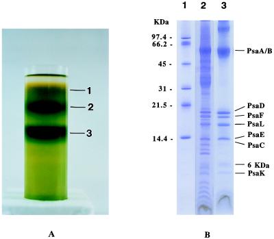
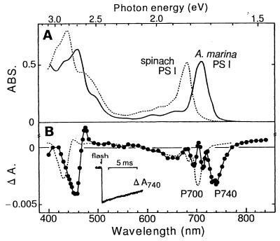
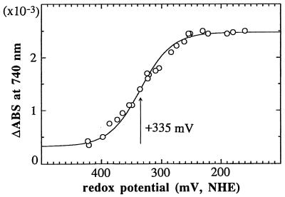
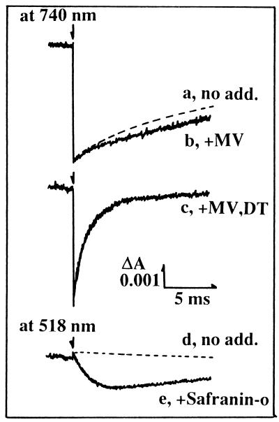
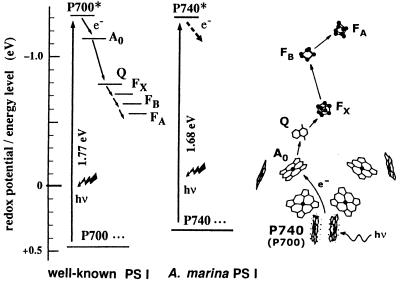
A far-red type of oxygenic photosynthesis was discovered in Acaryochloris marina, a recently found marine prokaryote that produces an atypical pigment chlorophyll d (Chl d). The purified photosystem I reaction center complex of A. marina contained 180 Chl d per 1 Chl a with PsaA-F, -L, -K, and two extra polypeptides. Laser excitation induced absorption changes of reaction center Chl d that was named P740 after its peak wavelength. A midpoint oxidation reduction potential of P740 was determined to be +335 mV. P740 uses light of significantly low quantum energy (740 nm = 1.68 eV) but generates a reducing power almost equivalent to that produced by a special pair of Chl a (P700) that absorbs red light at 700 nm (1.77 eV) in photosystem I of plants and cyanobacteria. The oxygenic photosynthesis based on Chl d might either be an acclimation to the far-red light environments or an evolutionary intermediate between the red-absorbing oxygenic and the far-red absorbing anoxygenic photosynthesis that uses bacteriochlorophylls.


Figures

Figure 1
Figure 1
Chemical structure of chlorophylls in oxygenic photosynthesis of plants and cyanobacteria.
Figure 2
Figure 2
(A) Separation of the pigment–protein complexes from detergent-solubilized thylakoid membranes of A. marina by sucrose density gradient centrifugation. The membranes were solubilized at 4°C for 30 min with 0.8% β-dodecyl maltoside, 20 mM Bis-Tris (pH 7.0), 10% glycerol, 10 mM NaCl, 10 mM CaCl2 and 2 mM EDTA-Na2. The sucrose gradient centrifugation (198,600 × g for 16 h) yielded three pigment-protein bands (1–3 on the right). (B) Polypeptide composition of the thylakoids and purified PS I complex from A. marina. Materials were treated with SDS and resolved on a 16–22% SDS/urea-PAGE. Lanes: 1, molecular weight standards; 2, thylakoid membranes; 3, PS I reaction center complex. The gel was stained with Coomassie blue. Sizes of molecular weight standards are indicated on the left, and subunit identifications based on N-terminal amino acid sequencing analysis are indicated on the right.
Figure 3
Figure 3
(A) Absorption (ABS) spectrum (Upper) and (B) laser flash-induced difference absorption (ΔA) spectrum (Lower) at 15°C of A. marina PS I complex (solid line, filled circles). The PS I complexes (band 3 in Fig. 2A) was suspended in Tris⋅HCl buffer (pH 7.5) containing 100 mM NaCl and 0.3 mM sodium ascorbate. Absorption and difference absorption spectra of P700 of spinach PS I (dashed line) are also shown.
Figure 4
Figure 4
Redox titration of the primary donor (P740) of A. marina PS I. The initial extent of the absorption change (ΔABS) at 740 nm after a laser flash excitation was plotted against the redox potential of the medium. The midpoint redox potential value was estimated to be +335 mV as shown by a fitting with an one-electron Nernst’s curve by assuming 5% irreversible absorption change (solid line).
Figure 5
Figure 5
Absorption changes of P740 and Safranin-o after laser flash excitation in A. marina PS I complex. Absorption changes were monitored at 740 nm in traces a–c and at 518 nm in traces d and e. Traces: a, no addition; b, with 300 μM methyl viologen; c, with 300 μM methyl viologen and 2 mM dithionite; d, no addition; e, with 40 μM Safranin-o. Other experimental conditions were similar to those in Fig. 3.
Figure 6
Figure 6
Electron transfer in the PS I reaction center. (Left) Electron transfer pathways in PS I of plants, cyanobacteria, and A. marina. Light induces the electron transfer from the donor (P700 in plants or cyanobacteria, and P740 in A. marina) to the acceptor chlorophyll (A0), quinone (Q), and the 4Fe4S clusters (FX, FB, and FA). The acceptor side of A. marina PS I is not known. (Right) Schematic representation of the central portion of PS I reaction center complex modified from the x-ray structure of cyanobacteria (16). In plants and cyanobacteria the chlorophylls including P700 and A0 are Chl a (9, 15, 16). In A. marina, P740 can be estimated to be a special pair of Chl d.

References

    1. Kok B. Plant Physiol. 1959;34:184–192. - PMC - PubMed
    1. Doering G, Renger G, Vater J, Witt H T. Z Naturforsch B. 1969;24:1139–1143. - PubMed
    1. Chisholm S W, Olson R J, Zettler E R, Goericke R, Waterbury J B, Welshmeyer N A. Nature (London) 1988;334:340–343.
    1. Miyashita H, Adachi K, Kurano N, Ikemoto H, Chihara M, Miyachi S. Nature (London) 1996;383:402.
    1. Miyashita H, Adachi K, Kurano N, Ikemoto H, Chihara M, Miyachi S. Plant Cell Physiol. 1997;38:274–281.

LinkOut - more resources