

Review
doi: 10.1128/JB.180.21.5495-5504.1998.
The Yersinia deadly kiss
Affiliations
- PMID: 9791096
- PMCID: PMC107605
- DOI: 10.1128/JB.180.21.5495-5504.1998
Item in Clipboard
Review
The Yersinia deadly kiss
J Bacteriol.
1998 Nov.
No abstract available
Figures
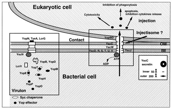
The basic model. When yersiniae are placed at 37°C in a rich environment, the Ysc secretion apparatus is installed and a stock of Yop proteins is synthesized. As long as there is no contact with a eukaryotic cell, a stop valve, possibly made of YopN, TyeA, and LcrG, blocks the Ysc secretion channel. Upon contact with the eukaryotic target cell, a sensor interacts with a receptor on the cell surface, which results in the opening of the secretion channel at the zone of contact. The Yops are then transported through the secretion channel, and the Yop effectors are translocated across the plasma membrane guided by YopB and -D. During their intrabacterial stage, Yops are capped with their specific chaperones, presumably to prevent premature associations. The rectangle on the left encloses the virulon, while that on the right encloses the injectisome.
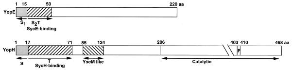
Schematic representation of YopE and YopH. S1, first secretion domain; S2/T, second secretion domain and translocation domain; P, catalytic P-loop site. Residues 85 to 124 of YopH present a significant but unexplained similarity to the hypothetical regulator LcrQ/YscM. aa, amino acids.
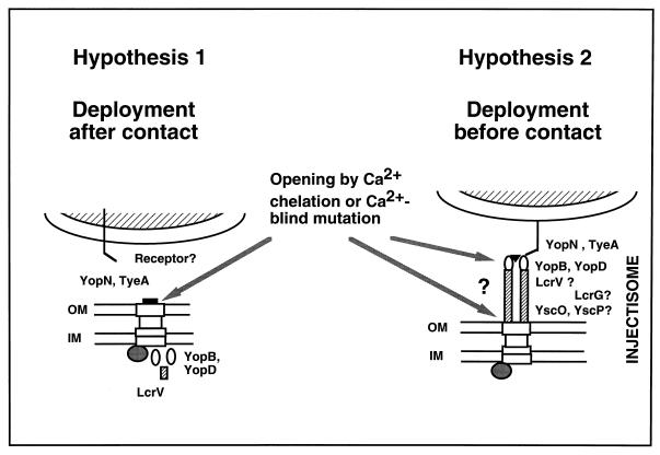
Two models for translocation. In the first hypothesis, the Ysc secretion apparatus (the syringe) is installed in the bacterial membranes but is closed by YopN and TyeA. Upon contact with a eukaryotic cell, the plug is removed and the translocation apparatus, composed of YopB, YopD, and LcrV (the needle?), grows into the eukaryotic cell. This model is supported by the observation that in vitro, the translocator Yops are secreted only upon Ca2+ chelation, like the other Yops. In this hypothesis, Ca2+ chelation would remove the YopN-TyeA plug, allowing secretion of all of the Yops. In the second hypothesis, the needle is installed before contact. This model is inspired by the electron microscopy observations of Kubori et al. (53) with S. typhimurium. In this model, Ca2+ chelation would either remove a cap at the tip of the needle or separate the needle from the basal body, leaving a large hole and inducing massive secretion of all of the Yops. LcrG could be a critical element at the base of the needle. OM, outer membrane; IM, inner membrane.
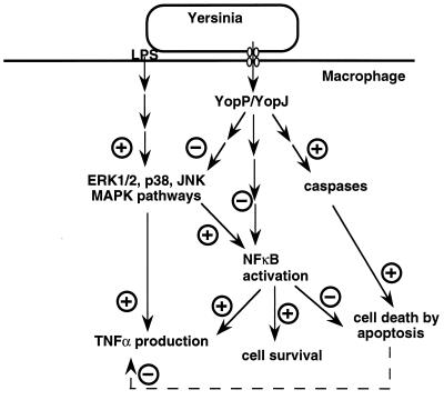
Model showing the effects of YopP/YopJ on the macrophage intracellular cascades. Lipopolysaccharide (LPS) activates the ERK1/2, JNK, and p38 MAPK pathways, which leads to increased TNF-α production. Activated MAPKs can lead to NFκB activation; activated NFκB can, in turn, enhance TNF-α transcription. Translocated YopP/YopJ induces macrophage apoptosis by a mechanism involving caspase activation. It also downregulates MAPKs and impairs NFκB activation, two effects that could explain the YopP/YopJ-induced reduction of TNF-α production. See the text for details and references.
References
-
- Allaoui A, Schulte R, Cornelis G R. Mutational analysis of the Yersinia enterocolitica virC operon: characterization of yscE, F, G, I, J, K required for Yop secretion and yscH encoding YopR. Mol Microbiol. 1995;18:343–355. - PubMed
-
- Anderson D M, Schneewind O. A mRNA signal for the type III secretion of Yop proteins by Yersinia enterocolitica. Science. 1997;278:1140–1143. - PubMed
Publication types
MeSH terms
Substances
LinkOut - more resources
Full Text Sources
Other Literature Sources
