. 1998 Nov;180(21):5580-90.
doi: 10.1128/JB.180.21.5580-5590.1998.

Domain analysis of the FliM protein of Escherichia coli

Affiliations

Domain analysis of the FliM protein of Escherichia coli

M A Mathews et al. J Bacteriol. 1998 Nov.

Abstract

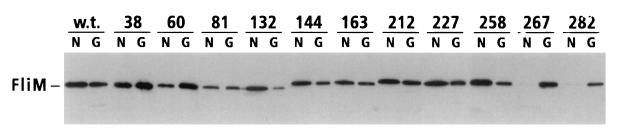
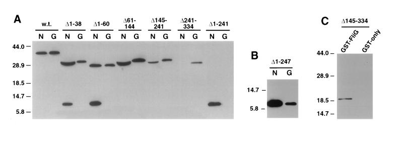
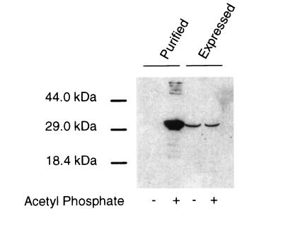
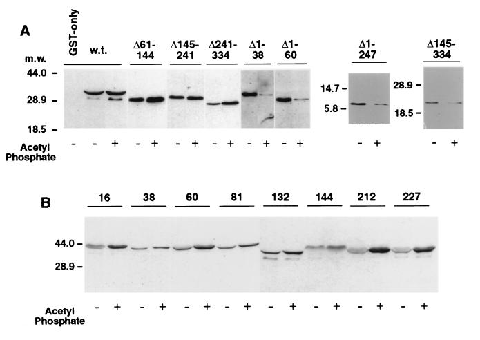
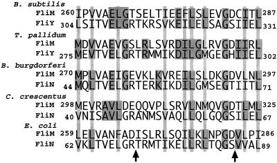
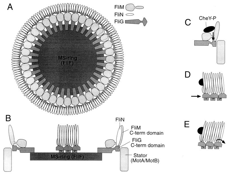
The FliM protein of Escherichia coli is required for the assembly and function of flagella. Genetic analyses and binding studies have shown that FliM interacts with several other flagellar proteins, including FliN, FliG, phosphorylated CheY, other copies of FliM, and possibly MotA and FliF. Here, we examine the effects of a set of linker insertions and partial deletions in FliM on its binding to FliN, FliG, CheY, and phospho-CheY and on its functions in flagellar assembly and rotation. The results suggest that FliM is organized into multiple domains. A C-terminal domain of about 90 residues binds to FliN in coprecipitation experiments, is most stable when coexpressed with FliN, and has some sequence similarity to FliN. This C-terminal domain is joined to the rest of FliM by a segment (residues 237 to 247) that is poorly conserved, tolerates linker insertion, and may be an interdomain linker. Binding to FliG occurs through multiple segments of FliM, some in the C-terminal domain and others in an N-terminal domain of 144 residues. Binding of FliM to CheY and phospho-CheY was complex. In coprecipitation experiments using purified FliM, the protein bound weakly to unphosphorylated CheY and more strongly to phospho-CheY, in agreement with previous reports. By contrast, in experiments using FliM in fresh cell lysates, the protein bound to unphosphorylated CheY about as well as to phospho-CheY. Determinants for binding CheY occur both near the N terminus of FliM, which appears most important for binding to the phosphorylated protein, and in the C-terminal domain, which binds more strongly to unphosphorylated CheY. Several different deletions and linker insertions in FliM enhanced its binding to phospho-CheY in coprecipitation experiments with protein from cell lysates. This suggests that determinants for binding phospho-CheY may be partly masked in the FliM protein as it exists in the cytoplasm. A model is proposed for the arrangement and function of FliM domains in the flagellar motor.


Figures

FIG. 1
FIG. 1
Coprecipitation of linker insertion mutant FliM proteins with GST-FliN (lanes labeled N) or GST-FliG (lanes labeled G). Positions of the linker insertions in FliM are indicated at the top. The experiment used the one-cell protocol (Materials and Methods). Coprecipitated material was analyzed on immunoblots probed with anti-FliM antiserum. Immunoblots of samples not exposed to the glutathione beads showed that all of the insertion mutant FliM proteins were present in cell lysates at levels comparable to that of the wild-type (w.t.) protein (data not shown).
FIG. 2
FIG. 2
Coprecipitation of FliM fragments with GST-FliN (lanes labeled N) and GST-FliG (lanes labeled G). The parts of FliM deleted are indicated at the top of each lane. (A) An initial set of FliM deletions, which together span the protein. The C-terminal fragment FliM242–334 (labeled Δ1-241 over the two rightmost lanes) was not stable except in the presence of FliN or GST-FliN, and so its failure to coprecipitate with GST-FliG is inconclusive. (B) Coprecipitation of an 87-residue C-terminal fragment of FliM with both GST-FliN and GST-FliG. This fragment accumulated in cells to detectable levels even in the absence of FliN (see text). (C) Coprecipitation of a 144-residue N-terminal fragment of FliM with GST-FliG. In negative-control experiments, neither FliM nor any of the FliM fragments was coprecipitated with GST alone (example control for a FliM fragment is shown here, and that for full-length FliM is shown in Fig. 4). w.t., wild type. Numbers to the left of each panel show molecular mass in kilodaltons.
FIG. 3
FIG. 3
Coprecipitation of FliM with GST-CheY in the presence or absence of agents that phosphorylate CheY, with FliM from two sources. (Left lanes) Experiment using FliM that was purified as described elsewhere (19, 32). Purified FliM was added to a suspension of RP3098 cells (which express no flagellar proteins) to give a final FliM level similar to that in other binding experiments. These FliM-supplemented cells were mixed with cells expressing GST-CheY, the cells were lysed, and a binding experiment was performed by the two-cell protocol. (Right lanes) A binding experiment done in the same way, except that the FliM was expressed within the RP3098 cells, and the samples were not supplemented with purified FliM.
FIG. 4
FIG. 4
Effects of deletions or linker insertions in FliM on binding to CheY in the presence or absence of the phosphorylating agent acetyl phosphate (all experiments contained Mg2+). (A) Coprecipitation of FliM deletion-mutant proteins with GST-CheY. The FliM deletions are indicated at the top. For the experiment using wild-type FliM protein, the GST-only negative control is also shown (first lane in panel A); all other lanes used the GST-CheY fusion protein. Negative controls for each of the FliM fragments showed that none was coprecipitated with GST alone (data not shown). Blots were typically exposed to film for 5 min after addition of the chemiluminescence reagents, but with the Δ1–38 and Δ1–60 mutants, the binding was somewhat weaker in the lanes without acetyl phosphate and significantly weaker in the lanes with acetyl phosphate, so overnight exposure was used. (The densitometry results in Table 3 used uniform exposures and a range of protein concentrations.) (B) Coprecipitation of FliM linker insertion proteins with GST-CheY. Positions of the linker insertions are indicated at the top. w.t., wild type. Numbers to the left of each panel show molecular mass (m.w.) in kilodaltons.
FIG. 5
FIG. 5
Patterns of sequence conservation, predicted secondary structures, and effects of linker insertions in FliM. The alignment used FliM sequences from E. coli, B. subtilis, B. burdorferi, and C. crescentus. The sequence from Agrobacterium tumefaciens is also known, but it is quite different from all of the others and was not included. Outlined and shaded boxes indicate residues that are identical in the four sequences, and nonoutlined, lightly shaded bars indicate positions where residues with hydrophobic character are found in all four sequences. Secondary structures were predicted by the neural net algorithm of Rost and Sander (21, 22), by using the information from all of the sequences. The α-helices are represented by coiled lines, the β-strands are represented by zigzag lines, and segments of nonregular secondary structure are represented by straight lines. Segments where predictions were ambiguous are left blank. Inverted triangles indicate positions and phenotypes of linker insertion mutations: solid, nonflagellate; stippled, flagellate but most cells nonmotile; striped, motile but nonswarming because of a strong CCW bias; and open, close to wild-type swarming. (See Table 2 for exact phenotypes and sequences of the insertions.)
FIG. 6
FIG. 6
Sequence alignments of segments of FliN with segments in the C-terminal domain of FliM, for species where both sequences are known (B. subtilis, Treponema pallidum, B. burgdorferi, C. crescentus, and E. coli). (The sequences are also known for Salmonella but are not significantly different from those of E. coli.) In B. subtilis and T. pallidum, the FliN homolog is called FliY and is a much larger protein that shows close homology to FliM in a short segment near the N terminus (1) (see the text). Residue numbers are not given for FliM from T. pallidum because the entire sequence is not known. Darkly shaded boxes indicate residues identical in FliM and FliN (or its homolog FliY) from the same species. Lightly shaded bars indicate positions where a sizable hydrophobic residue is found in both proteins from all species. Arrows indicate positions of the linker insertions in FliM that disrupted its binding to FliN.
FIG. 7
FIG. 7
Model of the arrangement of the FliG, FliM, and FliN proteins in the flagellar motor. (A) The FliG-FliM-FliN assembly mounted on the MS ring, as viewed from the cytoplasm. Subunit stoichiometries are approximate and are based on immunoblots of proteins in isolated flagellar structures (37, 38); FliN-FliM stoichiometries of 2:1 or 4:1 are also possible. The location of FliN in the C ring is based on immunoelectron microscopy (5, 38). Results of the present study suggest that FliN and the C-terminal domain of FliM occupy similar positions in the structure. The C-terminal domain of FliG is placed at the rotor-stator interface on the basis of mutational studies of FliG and of the stator proteins MotA and MotB (6, 7, 12, 13, 39, 40). (B) Side view of the FliG-FliM-FliN assembly. For clarity, only a subset of the proteins is shown. The cytoplasm is toward the top, and the periplasm is toward the bottom. The MS ring and MotA-MotB complexes are located in the cytoplasmic membrane, which is not pictured. (C to E) Hypotheses for the movements that might be triggered by binding of phospho-CheY to FliM, to cause switching to the CW direction of motor rotation. In each case, switching is suggested to involve a change in the position or orientation of the FliG C-terminal domain, relative to the stator and/or other parts of the rotor. (C) Binding of phospho-CheY might cause a domain of FliM, and the attached domain of FliG, to move up or down in a direction parallel to the rotation axis of the motor. (D) Phospho-CheY might induce subunits of the C ring to tilt, causing attached domains of FliG to move tangentially relative to other components of the rotor. Here and in panel E, the view is rotated 90° relative to that in panel C. (E) Phospho-CheY might induce tilting of both the C-ring subunits and the attached FliG domains, changing the angular orientation of the FliG domains.

Similar articles

Cited by

References

    1. Bischoff D S, Ordal G W. Identification and characterization of FliY, a novel component of the Bacillus subtilis flagellar switch complex. Mol Microbiol. 1992;6:2715–2723. - PubMed
    1. Bren A, Eisenbach M. The N-terminus of the flagellar switch protein, FliM, is the binding domain of the chemotactic response regulator, CheY. J Mol Biol. 1998;278:507–514. - PubMed
    1. Bren A, Welch M, Blat Y, Eisenbach M. Signal termination in bacterial chemotaxis: CheZ mediates dephosphorylation of free rather than switch-bound CheY. Proc Natl Acad Sci USA. 1996;93:10090–10093. - PMC - PubMed
    1. Francis N R, Irikura V M, Yamaguchi S, DeRosier D J, Macnab R M. Localization of the Salmonella typhimurium flagellar switch protein FliG to the cytoplasmic M-ring face of the basal body. Proc Natl Acad Sci USA. 1992;89:6304–6308. - PMC - PubMed
    1. Francis N R, Sosinsky G E, Thomas D, DeRosier D J. Isolation, characterization and structure of bacterial flagellar motors containing the switch complex. J Mol Biol. 1994;235:1261–1270. - PubMed

Publication types

LinkOut - more resources