. 1998 Nov;180(21):5652-9.
doi: 10.1128/JB.180.21.5652-5659.1998.

Identification, purification, and characterization of transpeptidase and glycosyltransferase domains of Streptococcus pneumoniae penicillin-binding protein 1a

Affiliations

Identification, purification, and characterization of transpeptidase and glycosyltransferase domains of Streptococcus pneumoniae penicillin-binding protein 1a

A M Di Guilmi et al. J Bacteriol. 1998 Nov.

Abstract

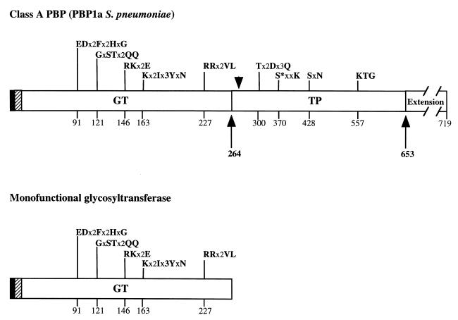
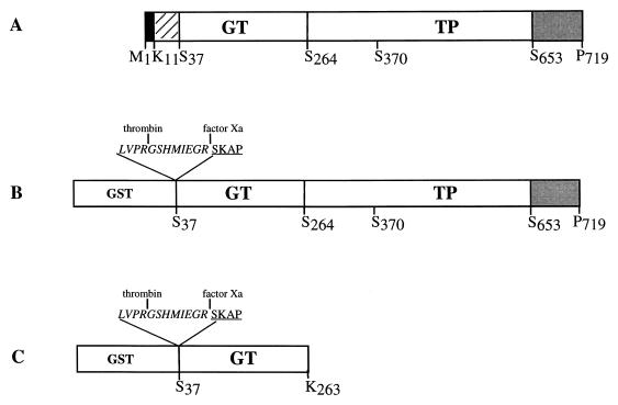
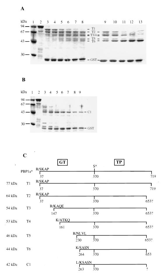
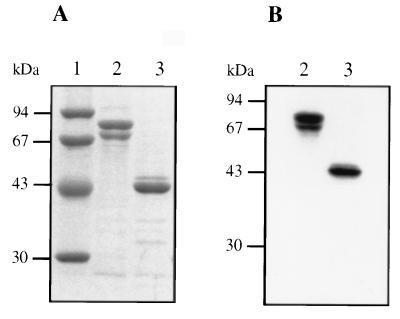
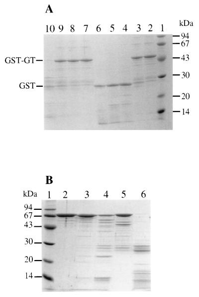
Resistance to beta-lactam antibiotics in Streptococcus pneumoniae is due to alteration of penicillin-binding proteins (PBPs). S. pneumoniae PBP 1a belongs to the class A high-molecular-mass PBPs, which harbor transpeptidase (TP) and glycosyltransferase (GT) activities. The GT active site represents a new potential target for the generation of novel nonpenicillin antibiotics. The 683-amino-acid extracellular region of PBP 1a (PBP 1a*) was expressed in Escherichia coli as a GST fusion protein. The GST-PBP 1a* soluble protein was purified, and its domain organization was revealed by limited proteolysis. A protease-resistant fragment spanning Ser 264 to Arg 653 exhibited a reactivity profile against both beta-lactams and substrate analogues similar to that of the parent protein. This protein fragment represents the TP domain. The GT domain (Ser 37 to Lys 263) was expressed as a recombinant GST fusion protein. Protection by moenomycin of the GT domain against trypsin degradation was interpreted as an interaction between the GT domain and the moenomycin.


Figures

FIG. 1
FIG. 1
Comparison of the molecular organization of class A PBP and MGT. The filled and hatched boxes represent the N-terminal cytoplasmic region and the membrane anchor, respectively. The C-terminal Ser- and Asn-rich extension is also depicted in PBP 1a. GT and TP domains are represented together with their conserved motifs; active serine 370 is indicated by an asterisk. x represents any amino acid. The amino acid numbering is that of S. pneumoniae PBP 1a (19). The arrows delineate the proteolytic PBP 1a* TP domain. The arrowhead refers to the permissive site for insertion mutations in E. coli PBP 1b as described by Lefèvre et al. (16).
FIG. 2
FIG. 2
Schematic diagrams of the PBP 1a-derived constructs. (A) Organization of the native PBP 1a protein. Filled, hatched, and shaded boxes indicate the N-terminal cytoplasmic region, the membrane anchor, and the Ser- and Asn-rich C-terminal extension, respectively. (B) Construction of the GST-PBP 1a* fusion protein. (C) Construction of the GST-GT fusion protein. The active-site serine 370 is indicated. Peptide at the GST-GT junctions includes sequences specific for thrombin and factor Xa (italic characters) and N-terminal amino acids of GT (underlined characters).
FIG. 3
FIG. 3
Analysis of purified GST-PBP 1a* and PBP 1a*. (A) Proteins were separated by SDS–12.5% PAGE and stained with Coomassie blue. (B) Autoradiogram of a dried SDS–12.5% PAGE of proteins (2 μg) labelled with [3H]benzylpenicillin prior to the migration. Numbers at the left indicate sizes of standard molecular mass markers. Lanes: 1, molecular mass markers; 2, GST-PBP 1a*; 3, PBP 1a*.
FIG. 4
FIG. 4
Characterization of peptides derived from limited proteolysis of GST-PBP 1a*. (A) Trypsin digestion. Lanes: 1, standard molecular mass markers; 2 to 8, incubation times of 15, 30, 60, 90, 120, and 150 min, respectively, at a trypsin/GST-PBP 1a* ratio of 1:8 (wt/wt); 9 to 13, incubation times of 30, 60, 120, 180, and 240 min, respectively, at a trypsin/GST-PBP 1a* ratio of 1:4 (wt/wt). (B) Chymotrypsin digestion. Lanes: 1, standard molecular mass markers; 2 to 9, incubation time of 5, 15, 30, 60, 90, 120, and 150 min, respectively, at a chymotrypsin/GST-PBP 1a* ratio of 1:8 (wt/wt). Numbers at the left indicate sizes of standard molecular mass markers. Protein fragments and GST are identified. (C) Schematic representation of the proteolytic fragments. The molecular mass of each fragment was measured by SDS-PAGE. The N-terminal sequence of the fragments was experimentally determined. The putative C termini of T2, T3, T4, and T5 are shown. The C terminus of T6 is derived from mass spectrometry measurements. The positions of the active-site serine 370 and the GT and TP domains are indicated. K/ and R/, trypsin digestion sites; L/, chymotrypsin site (the number corresponds to the N-terminal residue freed after digestion).
FIG. 5
FIG. 5
Analysis of purified PBP 1a* and TP. (A) Proteins were separated by SDS–12.5% PAGE and stained with Coomassie blue. (B) Autoradiogram of a dried SDS–12.5% PAGE of proteins (2 μg) labelled with [3H]benzylpenicillin prior to the migration. Numbers at the left indicate sizes of standard molecular mass markers. Lanes: 1, standard molecular mass markers; 2, PBP 1a*; 3, TP.
FIG. 6
FIG. 6
(A) Protection of GST-GT from trypsin hydrolysis by moenomycin. Lanes: 1, standard molecular mass markers; 2, no trypsin; 3 and 7, trypsin/GST-GT ratio of 1:1,500 (wt/wt); 4 and 8, trypsin/GST-GT ratio of 1:150 (wt/wt); 5 and 9, trypsin/GST-GT ratio of 1:75 (wt/wt); 6 and 10, trypsin/GST-GT ratio of 1:15 (wt/wt). Lanes 7 to 10, hydrolysis in the presence of 5 mM moenomycin. Numbers at the left indicate sizes of standard molecular mass markers. (B) Trypsin digestion of PBP 2x. Lanes: 1, molecular mass standards; 2, no trypsin; 3 and 5, trypsin/PBP 2x ratio of 1:100 (wt/wt) 4 and 6, trypsin/PBP 2x ratio of 1:10 (wt/wt). Lanes 5 and 6, hydrolysis in the presence of 5 mM moenomycin.

References

    1. Di Berardino M, Dijkstra A, Stüber D, Keck W, Gubler M. The monofunctional glycosyltransferase of Escherichia coli is a member of a new class of peptidoglycan-synthesising enzymes. Overexpression and determination of the glycan-polymerisation activity. FEBS Lett. 1996;392:184–188. - PubMed
    1. Dowson C G, Hutchison A, Brannigan J A, George R C, Hansman D, Linares J, Tomasz A, Maynard Smith J, Spratt B G. Horizontal transfer of penicillin-binding protein genes in penicillin resistant clinical isolates of Streptococcus pneumoniae. Proc Natl Acad Sci USA. 1989;86:8842–8846. - PMC - PubMed
    1. Dowson C G, Hutchison A, Woodford N, Johnson A P, George R C, Spratt B G. Penicillin-resistant viridans streptococci have obtained altered penicillin-binding protein genes from penicillin-resistant strains of Streptococcus pneumoniae. Proc Natl Acad Sci USA. 1990;87:5858–5862. - PMC - PubMed
    1. Dowson C G, Coffey T J, Spratt B G. Origin and molecular epidemiology of penicillin-binding protein-mediated resistance to β-lactam antibiotics. Trends Microbiol. 1994;2:361–366. - PubMed
    1. Georgopapadakou N H. Penicillin-binding proteins and bacterial resistance to β-lactams. Antimicrob Agents Chemother. 1993;37:2045–2053. - PMC - PubMed

Publication types

MeSH terms