

Monoclonal antibodies raised against native major capsid proteins of lactococcal c2-like bacteriophages
- PMID: 9797273
- PMCID: PMC106635
- DOI: 10.1128/AEM.64.11.4255-4259.1998
Monoclonal antibodies raised against native major capsid proteins of lactococcal c2-like bacteriophages
Abstract
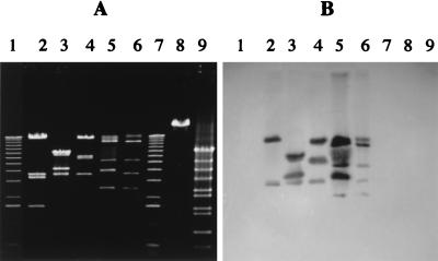
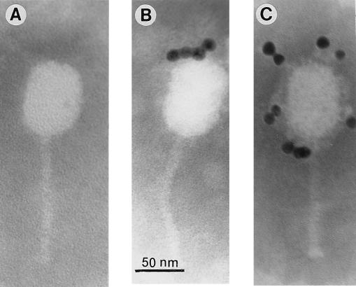
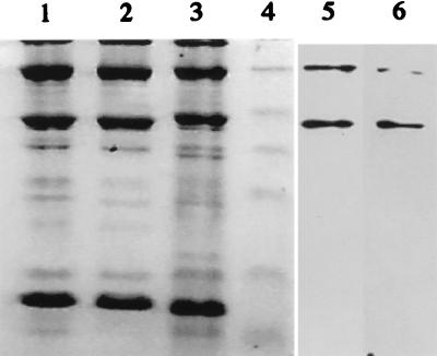
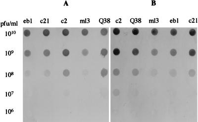
Phage Q38, a representative member of the c2 species, was purified by CsCl gradient and used to immunize BALB/c mice. Monoclonal antibodies (MAbs) were raised and then characterized by enzyme-linked immunosorbent assay. Two MAbs of isotype immunoglobulin G2a, designated 2A5 and 6G7, reacted only with phages belonging to the c2 species and not with phages of the 936 and P335 species, with a Lactococcus lactis cell extract, or with phage DNA. Immunoelectron microscopy showed that both MAbs recognized only phage head proteins. They did not react with any denatured phage proteins in Western blot assays. However, when the nitrocellulose membranes were treated with a Triton-based buffer to assist in protein renaturation, MAbs 2A5 and 6G7 recognized the two major capsid proteins with molecular masses of 80 and 170 kDa. Competitive inhibition tests showed that the two MAbs bind to overlapping epitopes. These MAbs may be a useful tool for monitoring c2 bacteriophages during dairy fermentation and in genetic studies.
Figures
References
-
- Ackermann H W, Dubow M S. Viruses of prokaryotes. Vol. 1. Boca Raton, Fla: CRC Press; 1987.
-
- Braun V, Hertwig S, Neve H, Geis A, Teuber M. Taxonomic differentiation of bacteriophages of Lactococcus lactis by electron microscopy, DNA-DNA hybridization and protein profiles. J Gen Microbiol. 1989;135:2551–2560.
-
- Candlish A A G. Immunological methods in food microbiology. Food Microbiol. 1991;8:1–14. - PubMed
Publication types
MeSH terms
Substances
LinkOut - more resources
Full Text Sources
Miscellaneous
