Comparative Study
. 1998 Nov;64(11):4446-51.
doi: 10.1128/AEM.64.11.4446-4451.1998.

Comparison of the thermostability properties of three acid phosphatases from molds: Aspergillus fumigatus phytase, A. niger phytase, and A. niger PH 2.5 acid phosphatase

Affiliations
Comparative Study

Comparison of the thermostability properties of three acid phosphatases from molds: Aspergillus fumigatus phytase, A. niger phytase, and A. niger PH 2.5 acid phosphatase

M Wyss et al. Appl Environ Microbiol. 1998 Nov.

Abstract

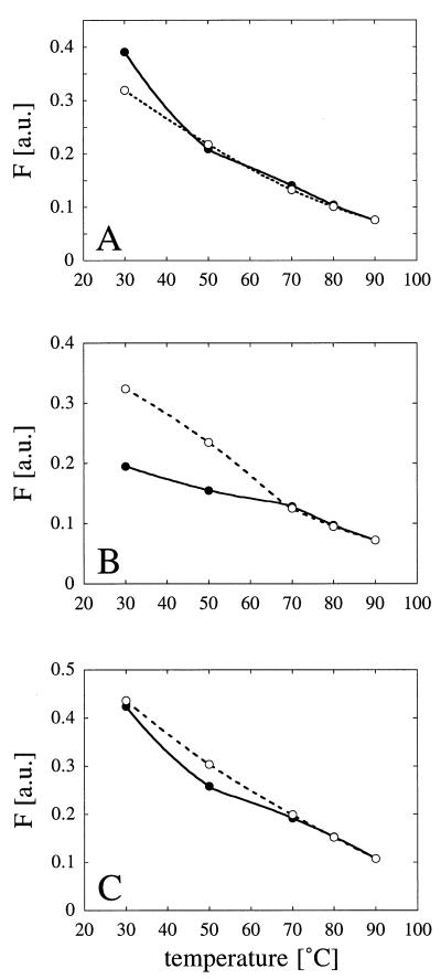
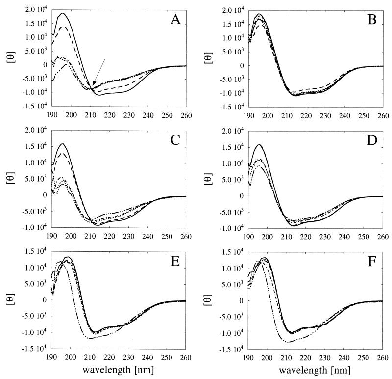
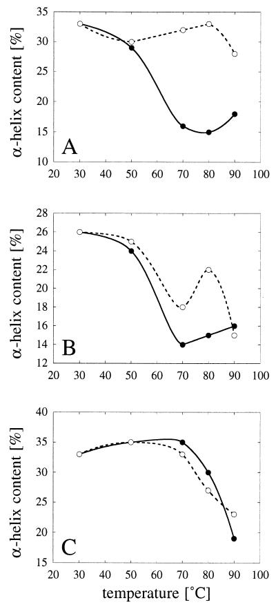
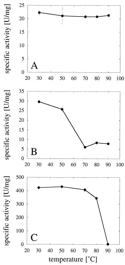
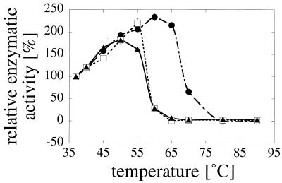
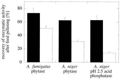
Enzymes that are used as animal feed supplements should be able to withstand temperatures of 60 to 90 degrees C, which may be reached during the feed pelleting process. The thermostability properties of three histidine acid phosphatases, Aspergillus fumigatus phytase, Aspergillus niger phytase, and A. niger optimum pH 2.5 acid phosphatase, were investigated by measuring circular dichroism, fluorescence, and enzymatic activity. The phytases of A. fumigatus and A. niger were both denatured at temperatures between 50 and 70 degrees C. After heat denaturation at temperatures up to 90 degrees C, A. fumigatus phytase refolded completely into a nativelike, fully active conformation, while in the case of A. niger phytase exposure to 55 to 90 degrees C was associated with an irreversible conformational change and with losses in enzymatic activity of 70 to 80%. In contrast to these two phytases, A. niger pH 2.5 acid phosphatase displayed considerably higher thermostability; denaturation, conformational changes, and irreversible inactivation were observed only at temperatures of >/=80 degrees C. In feed pelleting experiments performed at 75 degrees C, the recoveries of the enzymatic activities of the three acid phosphatases were similar (63 to 73%). At 85 degrees C, however, the recovery of enzymatic activity was considerably higher for A. fumigatus phytase (51%) than for A. niger phytase (31%) or pH 2.5 acid phosphatase (14%). These findings confirm that A. niger pH 2.5 acid phosphatase is irreversibly inactivated at temperatures above 80 degrees C and that the capacity of A. fumigatus phytase to refold properly after heat denaturation may favorably affect its pelleting stability.


Figures

FIG. 1
FIG. 1
Temperature-dependent changes in protein fluorescence. (A) A. fumigatus phytase. (B) A. niger T213 phytase. (C) A. niger pH 2.5 acid phosphatase. Symbols: •, fluorescence changes associated with increases in temperature from 30 to 50, 70, 80, and 90°C; ○, fluorescence changes associated with decreases in temperature from 90 to 80, 70, 50, and 30°C. F, fluorescence; a.u., arbitrary units. For experimental details see Materials and Methods.
FIG. 2
FIG. 2
Temperature-dependent unfolding and refolding of phytases and pH 2.5 acid phosphatase as determined by CD measurements. (A and B) A. fumigatus phytase. (C and D) A. niger T213 phytase. (E and F) A. niger pH 2.5 acid phosphatase. The individual acid phosphatases were incubated for 20 min at 30°C (——), 50°C (––––), 70°C (······), 80°C (–·–·–), or 90°C (–···–···–). Then either the CD spectra were determined directly at the same temperatures (A, C, and E), or the phosphatases were allowed to renature for 1 h at 30°C, and then the spectra were determined at 30°C (B, D, and F). The isodichroitic point observed for A. fumigatus phytase is indicated by an arrow in panel A. θ, residual molar ellipticity.
FIG. 3
FIG. 3
Temperature-dependent changes in calculated α-helical contents. (A) A. fumigatus phytase. (B) A. niger T213 phytase. (C) A. niger pH 2.5 acid phosphatase. The α-helical contents were calculated from the CD spectra shown in Fig. 2. Symbols: •, α-helical contents after 20 min of incubation at 30, 50, 70, 80, or 90°C; ○, α-helical contents after a subsequent 1-h renaturation period at 30°C.
FIG. 4
FIG. 4
Temperature-dependent enzyme inactivation. (A) A. fumigatus phytase. (B) A. niger T213 phytase. (C) A. niger pH 2.5 acid phosphatase. The individual acid phosphatases were incubated for 20 min at 30, 50, 70, 80, or 90°C and then immediately put on ice. The results represent means ± standard deviations (n = 3). Enzymatic activities were measured as described in Materials and Methods.
FIG. 5
FIG. 5
Effect of heat denaturation on the molecular sizes of phytases and pH 2.5 acid phosphatase. (A) A. fumigatus phytase. (B) A. niger T213 phytase. (C) A. niger pH 2.5 acid phosphatase. The individual acid phosphatases were incubated for 20 min at 30°C (curve a in panels A and B), 70°C (curve a in panel C), 80°C (curve b in panel C), and 90°C (curve b in panels A and B and curve c in panel C), allowed to refold for 2 h on ice, and then analyzed by gel permeation chromatography. The reference proteins blue dextran 2000 (Mr, 2,000,000), ferritin (Mr, 440,000), and albumin (Mr, 67,000) eluted at 8.5, 11.3, and 14.4 ml, respectively. OD280, optical density at 280 nm.
FIG. 6
FIG. 6
Temperature dependence of enzymatic activity. Symbols: ▴, A. fumigatus phytase; □, A. niger T213 phytase; •, A. niger pH 2.5 acid phosphatase. The enzymatic activities were measured directly at the temperatures indicated. The activity at 37°C was defined as 100%.
FIG. 7
FIG. 7
Recovery of enzymatic activity after feed pelleting. The three acid phosphatases were added in liquid form to extruded mash feed which was then pelleted at 75°C (solid bars) or 85°C (open bars). Phytase and pH 2.5 acid phosphatase activities were measured in the feed samples before and after pelleting.

References

    1. Chen G C, Yang J T. Two-point calibration of circular dichrometer with d-10-camphorsulfonic acid. Anal Lett. 1977;10:1195–1207.
    1. Delboni L F, Mande S C, Rentier-Delrue F, Mainfroid V, Turley S, Vellieux F M D, Martial J A, Hol W G J. Crystal structure of recombinant triosephosphate isomerase from Bacillus stearothermophilus. An analysis of potential thermostability factors in six isomerases with known three-dimensional structures points to the importance of hydrophobic interactions. Protein Sci. 1995;4:2594–2604. - PMC - PubMed
    1. Jobin Yvon. Instrument manual. Longjumeau, France: Jobin Yvon; 1990.
    1. Kawamura S, Kakuta Y, Tanaka I, Hikichi K, Kuhara S, Yamasaki N, Kimura M. Glycine-15 in the bend between two α-helices can explain the thermostability of DNA binding protein HU from Bacillus stearothermophilus. Biochemistry. 1996;35:1195–1200. - PubMed
    1. Kostrewa, D. Unpublished data.

Publication types

LinkOut - more resources