. 1998 Nov;5(6):773-9.
doi: 10.1128/CDLI.5.6.773-779.1998.

Antigenic structure of the nucleocapsid protein of porcine reproductive and respiratory syndrome virus

Affiliations

Antigenic structure of the nucleocapsid protein of porcine reproductive and respiratory syndrome virus

S K Wootton et al. Clin Diagn Lab Immunol. 1998 Nov.

Abstract

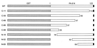
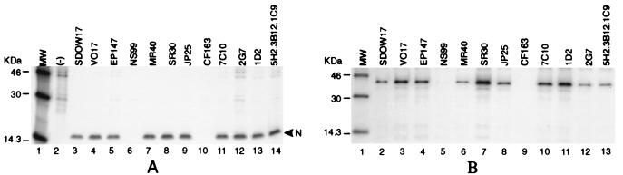
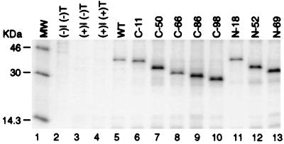
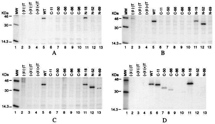
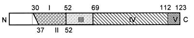
A collection of 12 monoclonal antibodies (MAbs) raised against porcine reproductive and respiratory syndrome (PRRS) virus was used to study the antigenic structure of the virus nucleocapsid protein (N). The full-length N gene, encoded by open reading frame 7, was cloned from the Canadian PRRS virus, PA-8. Deletions were introduced into the N gene to produce a series of nine overlapping protein fragments ranging in length from 25 to 112 amino acids. The individual truncated genes were cloned as glutathione S-transferase fusions into a eukaryotic expression vector downstream of the T7 RNA polymerase promoter. HeLa cells infected with recombinant vaccinia virus expressing T7 RNA polymerase were transfected with plasmid DNA encoding the N protein fragments, and the antigenicity of the synthesized proteins was analyzed by immunoprecipitation. Based on the immunoreactivities of the N protein deletion mutants with the panel of N-specific MAbs, five domains of antigenic importance were identified. MAbs SDOW17, SR30, and 5H2.3B12.1C9 each identified independent domains defined by amino acids 30 to 52, 69 to 123, and 37 to 52, respectively. Seven of the MAbs tested specifically recognized the local protein conformation formed in part by the amino acid residues 52 to 69. Furthermore, deletion of 11 amino acids from the carboxy terminus of the nucleocapsid protein disrupted the epitope configuration recognized by all of the conformation-dependent MAbs, suggesting that the carboxy-terminal region plays an important role in maintaining local protein conformation.


Figures

FIG. 1
FIG. 1
Schematic presentation of the N protein deletion mutants. The deletions and truncations of the N protein were constructed as described in Materials and Methods. Locations of the mutant proteins relative to the N protein are indicated by positions of amino acids. Shaded areas represent the GST coding sequence, and the black areas represent the translation termination.
FIG. 2
FIG. 2
(A) Specificity of the MAbs for the N protein of PA-8 virus. MARC-145 cells were infected with the PA-8 strain of PRRS virus at an MOI of 5 PFU/cell. Virus-infected cells were labelled with [35S]methionine at 50 μCi/ml, and cell extracts were prepared 48 h postinfection. Immunoprecipitation was performed with each of the 12 N-specific MAbs. Immune complexes were collected with protein A-Sepharose and resolved by SDS–12% PAGE as described in Materials and Methods. Uninfected cell lysate precipitated with a mixture of all 12 MAbs on the panel was used as a negative control (lane 2). The band migrating at 15 kDa representing the PA-8 N protein is marked with an arrowhead. (B) Specificity of the MAbs toward the recombinant GST-N fusion protein. HeLa cells infected with vaccinia virus expressing T7 RNA polymerase were transfected with plasmid DNA encoding the GST-N fusion protein under the control of the T7 promoter. Radiolabelled cell lysates were collected and immunoprecipitated with individual MAbs. The protein band (lanes 2 to 13) migrating at a molecular mass of 41 kDa represents the GST-N fusion protein. MW, molecular mass markers.
FIG. 3
FIG. 3
Expression of the GST-N fusion constructs immunoprecipitated with anti-GST antibody. HeLa cells infected with vaccinia virus expressing T7 RNA polymerase (vTF7-3) were transfected with plasmid DNA. Cells labelled with [35S]methionine were collected 26 h postinfection, immunoprecipitated with anti-GST antibody, and resolved by SDS–12% PAGE. Lane 1, molecular mass markers (MW); lane 2, uninfected and untransfected cells; lane 3, vTF7-3-infected, untransfected cells; lane 4, vTF7-3-infected, control plasmid DNA (pCITE2a)-transfected cells; lanes 5 to 13, lysates from cells expressing each of the N fusion proteins.
FIG. 4
FIG. 4
Immunoprecipitation of the GST-N protein deletion mutants with individual MAbs. HeLa cells were infected with vTF7-3 and transfected with plasmids encoding the N protein deletion mutants. Cells were radiolabelled with [35S]methionine (50 μCi/ml) for 16 h. Cell lysates containing the cytoplasmic fraction were immunoprecipitated with each of the N-specific MAbs, and the immune complexes were resolved by SDS–12% PAGE followed by autoradiography. (A) Immunoprecipitation with MAb SDOW17; (B) immunoprecipitation with MAb EP147 (representative also of the results observed for MAbs VO17, MR40, JP25, 1D2, 2G7, and 7C10); (C) immunoprecipitation with MAb SR30; (D) immunoprecipitation with MAb 5H2.3B12.1C9. Lane 1, molecular mass markers (MW); lane 2, uninfected, untransfected HeLa cells; lane 3, vTF7-3-infected, untransfected cells; lane 4, vTF7-3-infected, control plasmid DNA (pCITE2a)-transfected cells; lanes 5 to 13, lysates of cells expressing each of the N protein mutants.
FIG. 5
FIG. 5
Illustration of the five antigenically important domains localized on the N protein. The shaded areas flanked by amino acid positions represent domains localized by the mapping studies.

References

    1. Benfield D A, Nelson E, Collins J E, Harris L, Goyal S M, Robinson D, Christianson W T, Morrison R B, Gorcyca D E, Chladek D W. Characterization of swine infertility and respiratory syndrome (SIRS) virus (isolate ATCC VR-2332) J Vet Diagn Investig. 1992;4:127–133. - PubMed
    1. Cavanagh D. Nidovirales: a new order comprising Coronaviridae and Arteriviridae. Arch Virol. 1997;142:629–633. - PubMed
    1. Collins J E, Benfield D A, Christianson W T, Harris J E, Hennings L, Shaw J C, Goyal D P, McCullough S M, Morrison S, Joo R B, Gorcyca H S, Chladek D W. Isolation of swine infertility and respiratory syndrome virus (isolate ATCC VR-2332) in North America and experimental reproduction of the disease in gnotobiotic pigs. J Vet Diagn Investig. 1992;4:117–126. - PubMed
    1. Conzelmann K, Visser N, van Woensel P, Thiel H. Molecular characterization of porcine reproductive and respiratory syndrome virus, a member of the arterivirus group. Virology. 1993;193:329. - PMC - PubMed
    1. Denac H, Tratschin J D, Hofmann M A. An indirect ELISA for the detection of antibodies against porcine reproductive and respiratory syndrome virus using recombinant nucleocapsid protein as antigen. J Virol Methods. 1997;65:169–181. - PubMed

Publication types

MeSH terms

Associated data

LinkOut - more resources