. 1998 Nov 15;18(22):9294-302.
doi: 10.1523/JNEUROSCI.18-22-09294.1998.

Depolarization stimulates initial calcitonin gene-related peptide expression by embryonic sensory neurons in vitro

Affiliations

Depolarization stimulates initial calcitonin gene-related peptide expression by embryonic sensory neurons in vitro

X Ai et al. J Neurosci. .

Abstract

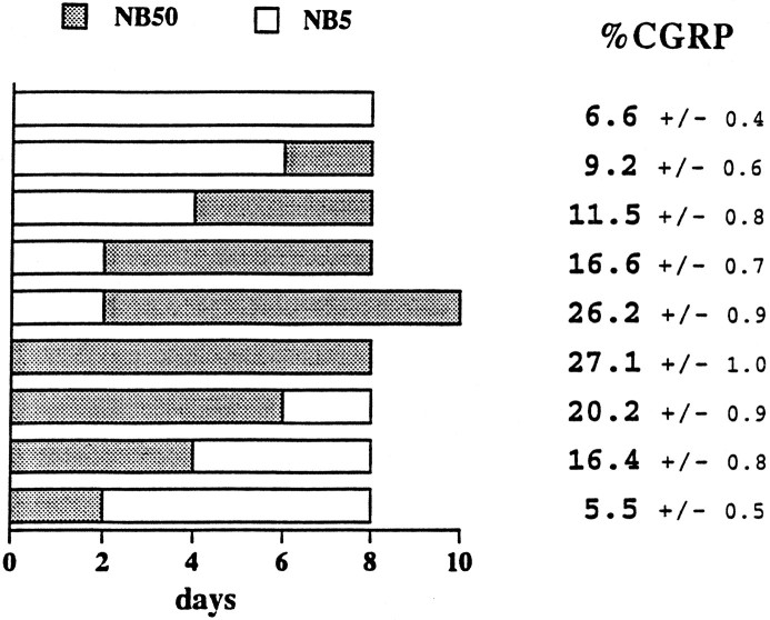
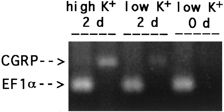
The neuropeptide calcitonin gene-related peptide (CGRP) is expressed by one-third of adult rat lumbar dorsal root ganglion (DRG) neurons, many of which mediate pain sensation or cause vasodilation. The factors that regulate the developmental expression of CGRP are poorly understood. Embryonic DRG neurons initially lack CGRP. When these neurons were stimulated in culture by serum or persistent 50 mM KCl application, the same percentage of CGRP-immunoreactive (CGRP-IR) neurons developed in vitro as was seen in the adult DRG in vivo. The addition of the L-type calcium channel blockers, 5 microM nifedipine or 10 microM verapamil, dramatically decreased the proportion of CGRP-IR neurons that developed, although the N-type calcium channel blocker, 2.5 microM omega-conotoxin, was less effective. By contrast, the sodium channel blocker 1 microM tetrodotoxin had no effect on CGRP expression after depolarization. Fura-2 ratiometric imaging demonstrated that mean intracellular free calcium levels increased from 70 to 135 nM with chronic depolarization, and the addition of nifedipine inhibited that increase. Only a subpopulation of neurons had elevated calcium concentrations during chronic depolarization, and they were correlated with CGRP expression. Key signal transduction pathways were tested pharmacologically for their role in CGRP expression after depolarization; the addition of the CaM kinase inhibitor KN-62 reduced the proportion of CGRP-IR neurons to basal levels. By contrast, protein kinase A and protein kinase C were not implicated in the depolarization-induced CGRP increases. These data suggest that depolarization and the subsequent Ca2+-based signal transduction mechanisms play important roles in the de novo expression of CGRP by specific embryonic DRG neurons.


Figures

Fig. 1.
Fig. 1.
CGRP expression was induced by serum or depolarization. Dissociated lumbar E14.5 DRGs were plated in basal defined NB5 medium containing 5 mm KCl or in depolarizing NB50 medium containing 50 mm KCl and compared with cultures in rat serum (RS). After 8 d the CGRP-IR neurons were quantified. A, The percentage of CGRP-IR neurons. In either depolarizing medium or in serum a similar proportion of neurons expressed CGRP, and the effects of depolarization and serum were not additive. By contrast, few neurons expressed CGRP in NB5 medium or in the iso-osmolarity control. Asterisksindicate the conditions under which CGRP-IR neurons were equivalent (p > 0.1) and that also differed from basal NB5 values (p < 0.0001). B, Cell survival was good in all conditions, with ∼80% of neurons alive after 8 d. Four independent experiments with each variable in triplicate were performed for A and B.C, CGRP immunocytochemistry in cultures with NB5 (5), NB50 (50), andRS. Few neurons in NB5 were lightly CGRP-immunoreactive (CGRP-IR). In NB50, some neurons had intense staining in the cell body and particularly in the perinuclear region. Intense CGRP staining was observed in the whole cell body of some neurons cultured inRS. D, The proportion of CGRP-IR neurons increased with increased KCl concentration. The concentration of KCl was varied from basal levels (NB5) to medium containing 75 mm KCl (NB75); CGRP-IR neurons were counted after 8 d in culture. Five or six independent experiments with each concentration were performed in triplicate. Approximately 85% of the neurons survived in each condition.
Fig. 2.
Fig. 2.
Maintained depolarization was required to elicit CGRP expression. Dissociated E14.5 DRG cells were maintained in NB5 or NB50. Growth media were switched at days 2, 4, or 6. After a total of 8 or 10 d the cultures were processed for CGRP immunocytochemistry. The proportion of CGRP-IR neurons was quantified, and the data represent the mean and SEM of eight independent experiments. The highest proportion of CGRP-IR neurons was seen with 8 d of depolarization (chronic or applied after 2 d in NB5 had the same results; p = 0.73). CGRP expression decreased if NB50 was removed during the culture period.
Fig. 3.
Fig. 3.
CGRP mRNA increased in DRG cultures with depolarization. Dissociated E14.5 DRG cells were placed in tissue culture in basal or depolarizing conditions. At the time indicated, total RNA was extracted and prepared for RT-PCR with primers specific for α-CGRP or the transcription factor elongation factor 1α (EF1α). In control cultures total RNA was extracted immediately (low K+, 0 d), and no CGRP mRNA was detected. After 2 d, CGRP mRNA was present in depolarizing (high K+, 2 d) and basal (low K+, 2 d) conditions. This result was obtained in three independent assays.
Fig. 4.
Fig. 4.
Calcium imaging revealed that DRG neurons maintained in depolarizing NB50 had elevated free intracellular calcium. A, Mean free intracellular calcium increased with depolarization and was reduced to basal levels after nifedipine treatment. Free intracellular Ca2+ levels were measured in neurons at day 4. DRG cells were grown for 4 d in NB5,NB50, or NB50 with 10 μm nifedipine before they were loaded with fura-2 AM. Free [Ca2+] inside the cell was quantified. At least 300 neurons from three independent experiments were analyzed for each condition.B–D, Ratio histograms. The distributions of free calcium values from neurons cultured in NB5, NB50, or NB50 with 10 μm nifedipine for 4 d are shown in histograms. Most neurons in NB50 had ratios similar to those in NB5, whereas a distinct subpopulation had “higher Ca2+” levels.E, Free calcium levels were higher in a subpopulation of neurons cultured in NB50 for 4 d, shown in the ratio measurement with light blue pseudocolor. Similar observations were obtained in six independent experiments.
Fig. 5.
Fig. 5.
Higher intracellular calcium levels were correlated with CGRP immunoreactivity. DRG cells were grown for 4 d in depolarizing NB50 on gridded coverslips, and free intracellular calcium was visualized by fura-2 fluorescence ratiometric imaging (A). Although many neurons had basal free calcium levels, some contained higher free calcium and appearedblue in pseudocolor (arrows). The cells were fixed and processed for CGRP immunocytochemistry (B), and the same neurons were located on gridded coverslips. Peptide expression was only just detectable at this early stage and appeared in a perinuclear, Golgi-like location in neurons. CGRP immunoreactivity was more likely to be found in neurons with elevated calcium (arrows).

Similar articles

Cited by

References

    1. Adler EM, Fink JS. Calcium regulation of vasoactive intestinal polypeptide mRNA abundance in SH-SY5Y human neuroblastoma cells. J Neurochem. 1993;61:727–737. - PubMed
    1. Bading H, Ginty DD, Greenberg ME. Regulation of gene expression in hippocampal neurons by distinct calcium signaling pathways. Science. 1993;260:181–186. - PubMed
    1. Bito H, Deisseroth K, Tsien RW. CREB phosphorylation and dephosphorylation: a Ca stimulus duration-dependent switch for hippocampal gene expression. Cell. 1996;87:1203–1214. - PubMed
    1. Bito H, Deisseroth K, Tsien RW. Ca2+-dependent regulation in neuronal gene expression. Curr Opin Neurobiol. 1997;7:419–429. - PubMed
    1. Brain SD, Williams TJ, Tippins JR, Morris HR, MacIntyre I. Calcitonin gene-related peptide is a potent vasodilator. Nature. 1985;313:54–56. - PubMed

Publication types

MeSH terms