Control of myelination by specific patterns of neural impulses
- PMID: 9801369
- PMCID: PMC6792896
- DOI: 10.1523/JNEUROSCI.18-22-09303.1998
Control of myelination by specific patterns of neural impulses
Abstract
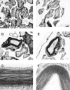
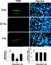
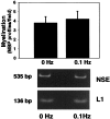
A cell culture preparation equipped with stimulating electrodes was used to investigate whether action potential activity can influence myelination of mouse dorsal root ganglia axons by Schwann cells. Myelination was reduced to one-third of normal by low-frequency impulse activity (0.1 Hz), but higher-frequency stimulation (1 Hz) had no effect. The number of Schwann cells and the ultrastructure of compact myelin were not affected. The frequency of stimulation that inhibited myelination decreased expression of the cell adhesion molecule L1, and stimulation under conditions that prevented the reduction in L1 blocked the effects on myelination. This link between myelination and functional activity in the axon at specific frequencies that change axonal expression of L1 could have important consequences for the structural and functional relationship of myelinating axons.
Figures


Similar articles
-
Inhibition of Schwann cell myelination in vitro by antibody to the L1 adhesion molecule.J Neurosci. 1990 Nov;10(11):3635-45. doi: 10.1523/JNEUROSCI.10-11-03635.1990. J Neurosci. 1990. PMID: 2230951 Free PMC article.
-
Nerve growth factor-hypersecreting Schwann cell grafts augment and guide spinal cord axonal growth and remyelinate central nervous system axons in a phenotypically appropriate manner that correlates with expression of L1.J Comp Neurol. 1999 Nov 1;413(4):495-506. doi: 10.1002/(sici)1096-9861(19991101)413:4<495::aid-cne1>3.0.co;2-z. J Comp Neurol. 1999. PMID: 10495438
-
Heterophilic binding of L1 on unmyelinated sensory axons mediates Schwann cell adhesion and is required for axonal survival.J Cell Biol. 1999 Sep 6;146(5):1173-84. doi: 10.1083/jcb.146.5.1173. J Cell Biol. 1999. PMID: 10477768 Free PMC article.
-
Schwann cell myelination.Cold Spring Harb Perspect Biol. 2015 Jun 8;7(8):a020529. doi: 10.1101/cshperspect.a020529. Cold Spring Harb Perspect Biol. 2015. PMID: 26054742 Free PMC article. Review.
-
Studies of the initiation of myelination by Schwann cells.Ann N Y Acad Sci. 1990;605:1-14. doi: 10.1111/j.1749-6632.1990.tb42376.x. Ann N Y Acad Sci. 1990. PMID: 2268113 Review.
Cited by
-
Brain white matter structure and COMT gene are linked to second-language learning in adults.Proc Natl Acad Sci U S A. 2016 Jun 28;113(26):7249-54. doi: 10.1073/pnas.1606602113. Epub 2016 Jun 13. Proc Natl Acad Sci U S A. 2016. PMID: 27298360 Free PMC article.
-
Adenosine: a neuron-glial transmitter promoting myelination in the CNS in response to action potentials.Neuron. 2002 Dec 5;36(5):855-68. doi: 10.1016/s0896-6273(02)01067-x. Neuron. 2002. PMID: 12467589 Free PMC article.
-
Neuronal activity in the hub of extrasynaptic Schwann cell-axon interactions.Front Cell Neurosci. 2013 Nov 25;7:228. doi: 10.3389/fncel.2013.00228. eCollection 2013. Front Cell Neurosci. 2013. PMID: 24324401 Free PMC article.
-
A suspended carbon fiber culture to model myelination by human Schwann cells.J Mater Sci Mater Med. 2017 Apr;28(4):57. doi: 10.1007/s10856-017-5867-x. Epub 2017 Feb 16. J Mater Sci Mater Med. 2017. PMID: 28210970
-
Neuronal Ndrg4 Is Essential for Nodes of Ranvier Organization in Zebrafish.PLoS Genet. 2016 Nov 30;12(11):e1006459. doi: 10.1371/journal.pgen.1006459. eCollection 2016 Nov. PLoS Genet. 2016. PMID: 27902705 Free PMC article.
References
-
- Aguayo AJ, Epps J, Charron L, Bray GM. Multipotentiality of Schwann cells in cross-anastomosed and grafted myelinated and unmyelinated nerves: quantitative microscopy and radioautography. Brain Res. 1976;104:1–20. - PubMed
-
- Barres BA, Raff MC. Proliferation of oligodendrocyte precursor cells depends on electrical activity in axons. Nature. 1993;361:258–260. - PubMed
-
- Carenini S, Montag D, Crener H, Schachner M, Martini R. Absence of the myelin-associated glycoprotein (MAG) and the neural cell adhesion molecule (N-CAM) interferes with the maintenance, but not with the formation of peripheral myelin. Cell Tissue Res. 1997;287:3–9. - PubMed
-
- Carenini S, Montag D, Schachner M, Martini R. MAG-deficient Schwann cells myelinate dorsal root ganglion neurons in culture. Glia. 1998;22:213–220. - PubMed
MeSH terms
Substances
LinkOut - more resources
Full Text Sources
