. 1998 Nov 15;18(22):9312-25.
doi: 10.1523/JNEUROSCI.18-22-09312.1998.

Neurotrimin mediates bifunctional effects on neurite outgrowth via homophilic and heterophilic interactions

Affiliations

Neurotrimin mediates bifunctional effects on neurite outgrowth via homophilic and heterophilic interactions

O D Gil et al. J Neurosci. .

Abstract

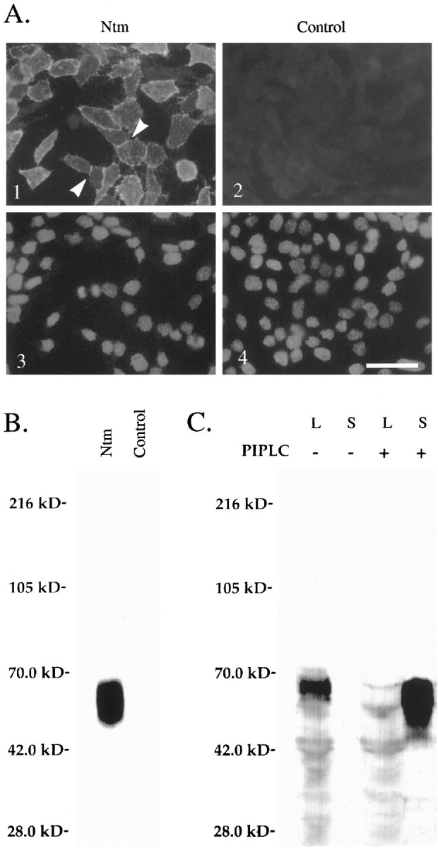
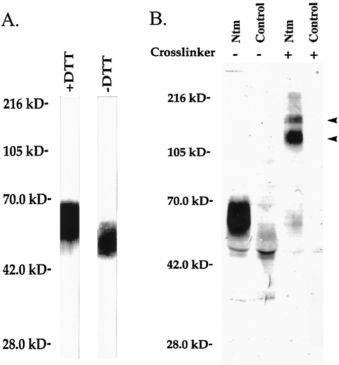
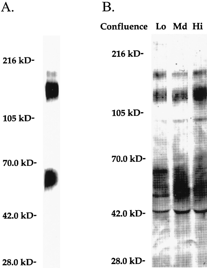
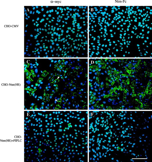
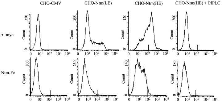
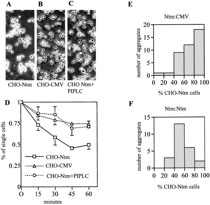
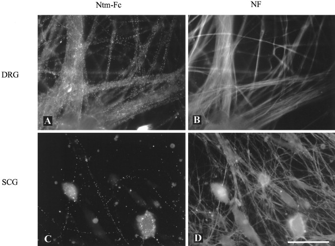
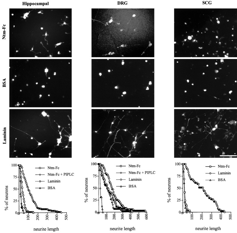
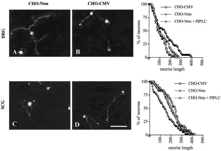
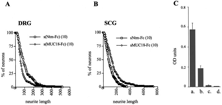
Neurotrimin (Ntm) together with the limbic system-associated membrane protein (LAMP) and the opioid-binding cell adhesion molecule (OBCAM) comprise the IgLON family of neural cell adhesion molecules. These glycosylphosphatidylinositol (GPI)-anchored proteins are expressed in distinct neuronal systems. In the case of Ntm, its expression pattern suggests a role in the development of thalamocortical and pontocerebellar projections (Struyket al., 1995). We have now characterized Ntm's function in cell adhesion and in neurite outgrowth. Cross-linking studies of transfected cells show that Ntm forms noncovalent homodimers and multimers at the cell surface. Ntm mediates homophilic adhesion, as evidenced by the reaggregation of the transfected cells and the specific binding of an Ntm-Fc chimera to these cells. Consistent with these results, Ntm-Fc binds to neurons that express Ntm at high levels, e.g., dorsal root ganglion (DRG) and hippocampal neurons. It does not bind to DRG neurons treated with phosphatidylinositol-specific phospholipase C (PI-PLC) or to sympathetic neurons that do not express Ntm or other members of the IgLON family at significant levels. Ntm promotes the outgrowth of DRG neurons, even after PI-PLC treatment, suggesting that its effects on outgrowth are mediated by heterophilic interactions. Of particular note, both membrane-bound and soluble Ntm inhibit the outgrowth of sympathetic neurons. These results strongly suggest that Ntm, and other members of the IgLON family, regulate the development of neuronal projections via attractive and repulsive mechanisms that are cell type specific and are mediated by homophilic and heterophilic interactions.


Figures

Fig. 1.
Fig. 1.
Characterization of Ntm-myc expression in transfected CHO cells. A, Immunofluorescence micrographs showing Ntm-myc expression on the surface of CHO-Ntm cells (panel 1) detected with an anti-myc antibody.Arrowheads indicate examples of the accumulation of Ntm at sites of cell–cell contact. Control cells transfected with the vector (panel 2) are not stained. Thebottom panels show the Hoechst staining of the corresponding fields. Scale bar, 50 μm. B, Lysates of surface-biotinylated CHO-Ntm and control cell monolayers were immunoprecipitated with the anti-myc antibody, electrophoresed, blotted, and probed with alkaline phosphatase-conjugated streptavidin. The prominent band migrating at 55–65 kDa, corresponding to the predicted size of Ntm, is present in immunoprecipitates of Ntm-myc cells but not control cells. Molecular weight markers are indicated atleft. C, Western blot, with the anti-myc antibody, of supernatants (S) and cell lysates (L) of CHO-Ntm cultures either treated (+) or mock-treated (−) with PI-PLC. A prominent band corresponding to Ntm-myc is present in the supernatant of PI-PLC-treated cultures but not in mock-treated cultures. Molecular weight markers are indicated atleft.
Fig. 2.
Fig. 2.
Ntm forms noncovalent homodimers.A, GPI-anchored proteins were extracted from confluent CHO-Ntm cells, electrophoresed under reducing (+DTT) or nonreducing (−DTT) conditions, blotted, and detected with phosphatase alkaline-conjugated streptavidin. Note that Ntm-myc is the only GPI-anchored protein expressed on the surface of CHO-Ntm cells and that there are no high molecular weight complexes under nonreducing conditions. Molecular weight markers are indicated atleft. B, Proteins were immunoprecipitated from surface biotinylated CHO-Ntm or CHO-CMV cell lysates with the anti-myc antibody after treatment (+) or mock-treatment (−) with BS3. In the BS3-treated CHO-Ntm cells, all of the Ntm-myc migrates in two high molecular weight complexes (arrowheads) with mobilities of 130 and 180 kDa. Molecular weight markers are indicated atleft.
Fig. 3.
Fig. 3.
Ntm forms oligomeric complexes at the cell surface. A, CHO-Ntm cells were treated with BS3, followed by PI-PLC treatment. The PI-PLC supernatant was electrophoresed and Western-blotted with the anti-myc antibody. The Ntm-myc cross-linked complexes appeared in the supernatant after PI-PLC release and were not cell-associated. Molecular weight markers are indicated at left.B, CHO-Ntm cells were plated at low, medium, and high densities, treated with BS3, and surface-biotinylated. Anti-myc antibody immunoprecipitates from cell lysates were electrophoresed, blotted, and probed with alkaline phosphatase-conjugated streptavidin. Ntm-myc cross-linked complexes were seen at both low and high densities, reflecting cisinteractions at the cell surface. Molecular weight markers are indicated at left.
Fig. 4.
Fig. 4.
Ntm mediates homophilic binding. Fields corresponding to control (A, B), CHO-Ntm (C, D), and CHO-Ntm cells pretreated with PI-PLC (E, F) were stained with the anti-myc antibody and FITC-conjugated anti-mouse IgG (A, C, E) or incubated with Ntm-Fc and FITC-conjugated anti-human Fc (B, D, F). Examples of the accumulation of Ntm at sites of cell–cell contact are marked with arrowheads(C). Scale bar, 100 μm.
Fig. 5.
Fig. 5.
Quantitation of Ntm-Fc binding by flow cytometry. The top row show FACS analysis of the expression of Ntm-myc by control (CHO-CMV),CHO-Ntm(LE), CHO-Ntm(HE), and CHO-Ntm(HE) cells pretreated with PI-PLC [CHO-NTM(HE) + PIPLC]. Analysis of the binding of Ntm-Fc to the same sequence of cells is shown in the bottom row. The abscissa corresponds to the log of fluorescence intensity.
Fig. 6.
Fig. 6.
Transfected CHO cells reaggregate by a homophilic mechanism. CHO cells (Ntm, control, and Ntm pretreated with PI-PLC) were dissociated and allowed to reaggregate for 60 min. Photomicrographs of representative aggregates that formed by Ntm-transfected CHO cells (A), control CHO cells (B), and Ntm-transfected CHO cells pretreated with PI-PLC (C) are shown. To examine the kinetics of reaggregation, aliquots were withdrawn at 15 min intervals, and single cells were counted in a hemocytometer (D). Values are the mean ± SEM for four independent experiments. Analysis of the composition of aggregates formed by a mixture of Ntm-transfected and control CHO cells demonstrates that aggregates are composed predominantly of Ntm-transfected cells (E). Control mixture of CHO-Ntm labeled with diI and diO reveals that the composition of aggregates is equally mixed (F).
Fig. 7.
Fig. 7.
Binding of Ntm-Fc to primary neurons. Cultures of dissociated DRG (A, B) and SCG neurons (C, D) are shown. Ntm-Fc binding (A, C) and neurofilament staining (B, D) are shown. Note that Ntm-Fc binds to the great majority of the DRG neurites but to only a few of the SCG neurites. Scale bar, 50 μm.
Fig. 8.
Fig. 8.
Ntm-Fc promotes hippocampal and DRG but not SCG neurite outgrowth. Photomicrographs show neurons cultured on 100 μg/ml Ntm-Fc, 1% BSA, or 10–25 μg/ml laminin adsorbed to nitrocellulose substrates. Hippocampal neurons (left column) were prelabeled with diI, whereas sensory (center column) and sympathetic neurons (right column) were stained with the neurofilament-specific antibody 3A10. Quantitation of the outgrowth for each cell type is shown in thegraphs at the bottom. Scale bar, 100 μm.
Fig. 9.
Fig. 9.
Ntm promotes DRG and inhibits SCG neurite outgrowth. Photomicrographs show DRG (A, B) and SCG (C, D) neurons cultured on a monolayer of Ntm-transfected CHO cells (A, C) or CHO cells transfected with the vector (B, D). Cultures were fixed and stained for neurofilament after an additional 14 hr.Graphs showing quantitation of neurite outgrowth for each cell type are on the right. Scale bar, 100 μm.
Fig. 10.
Fig. 10.
Soluble Ntm-Fc promotes DRG and inhibits SCG neurite outgrowth. Graphs show quantitation of DRG (A) and SCG (B) neurons plated on collagen and laminin substrates, respectively, in the presence of soluble forms of Ntm-Fc or MUC18-Fc. The abscissa corresponds to neurite length (in micrometers), and the ordinate corresponds to the percentage of neurons with neurites greater than a specific length. An ELISA is shown (C) in which Ntm-Fc was adsorbed to the plastic dish in buffer alone (a), in culture media with 1% BSA (b), or in the media but with the plastic precoated with collagen (c) or laminin (d).

References

    1. Benchimol S, Fuks A, Jothy S, Beauchemin N, Shirota K, Stanners CP. Carcinoembryonic antigen, a human tumor marker functions as an intercellular adhesion molecule. Cell. 1989;57:327–334. - PubMed
    1. Bixby JL, Harris WA. Molecular mechanisms of axon growth and guidance. Annu Rev Cell Biol. 1991;7:117–159. - PubMed
    1. Brewer GJ. Serum-free B27/neurobasal medium supports differentiated growth of neurons from the striatum, substantia nigra, septum, cerebral cortex, cerebellum, and dentate gyrus. J Neurosci Res. 1995;42:674–683. - PubMed
    1. Brieher WM, Yap AS, Gumbiner BM. Lateral dimerization is required for the homophilic binding activity of C-cadherin. J Cell Biol. 1996;135:487–496. - PMC - PubMed
    1. Brümmendorf T, Rathjen FG. Axonal glycoproteins with immunoglobulin- and fibronectin type III-related domains in vertebrates: structural features, binding activities, and signal transduction. J Neurochem. 1993;61:1207–1219. - PubMed

Publication types

MeSH terms