. 1998 Nov 15;18(22):9429-37.
doi: 10.1523/JNEUROSCI.18-22-09429.1998.

Sustained activity in the medial wall during working memory delays

Affiliations

Sustained activity in the medial wall during working memory delays

L Petit et al. J Neurosci. .

Abstract

We have taken advantage of the temporal resolution afforded by functional magnetic resonance imaging (fMRI) to investigate the role played by medial wall areas in humans during working memory tasks. We demarcated the medial motor areas activated during simple manual movement, namely the supplementary motor area (SMA) and the cingulate motor area (CMA), and those activated during visually guided saccadic eye movements, namely the supplementary eye field (SEF). We determined the location of sustained activity over working memory delays in the medial wall in relation to these functional landmarks during both spatial and face working memory tasks. We identified two distinct areas, namely the pre-SMA and the caudal part of the anterior cingulate cortex (caudal-AC), that showed similar sustained activity during both spatial and face working memory delays. These areas were distinct from and anterior to the SMA, CMA, and SEF. Both the pre-SMA and caudal-AC activation were identified by a contrast between sustained activity during working memory delays as compared with sustained activity during control delays in which subjects were waiting for a cue to make a simple manual motor response. Thus, the present findings suggest that sustained activity during working memory delays in both the pre-SMA and caudal-AC does not reflect simple motor preparation but rather a state of preparedness for selecting a motor response based on the information held on-line.


Figures

Fig. 1.
Fig. 1.
Working memory task diagram and description of the contrasts used for the multiple regression analysis. The contrasts made between task components are (1) visual stimulation versus no visual stimulation, (2) memory stimuli versus control stimuli, (3) the control stimulus set versus the control response, (4) the memory stimulus set versus the test stimulus and response, (5) delays during anticipation of response versus intertrial intervals, and (6) the memory delays versus the control delays (for details, see Materials and Methods) (adapted from Courtney et al., 1998b).
Fig. 2.
Fig. 2.
Color-coded Z-score map illustrating significant manual motor-related activity (top) (Fig. 1, regressor 3) and sustained activity during face working memory delays (bottom) (Fig. 1, regressor 6) in a single subject (S11), shown overlaid onto a Talairach normalized anatomical MR sagittal image (Y = −6 mm) for that subject with the cingulate sulcus shown inwhite. Note that the superior frontal and cingulate foci are anatomically distinct for both motor-related (top) and sustained (bottom) activity. AC-PC, Bicommissural plane; VAC and VPC, vertical planes passing through the anterior and posterior commissures, respectively.
Fig. 3.
Fig. 3.
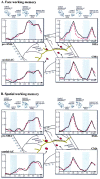
Plot of Talairach coordinates (in millimeters) of manual motor-related (red squares) and working memory delay-related activities during both face (top, red triangles) and spatial (bottom, red circles) working memory tasks (for details, see Tables 1, 2). Time series (inset graphs) illustrate the average response (in percentage change of MR signal, solid black line) and the corresponding fitted response function (dashed red line) from regression analysis. VAC, Vertical plane passing through the anterior commissure. The region of cortex inpink brain drawings is the same as that illustrated in the right part of Figure 4.
Fig. 4.
Fig. 4.
Left, Midsagittal view of the human brain that illustrates the VOIs defined in the present study, namely the dorsomedial part of the superior frontal cortex (orange) and the anterior and mid-cingulate cortex (dark blue) (for details, see Materials and Methods). Right, Plot of Talairach coordinates (in millimeters) of medial activations described in previous working memory studies in the medial superior frontal cortex (white filled circles) and in the cingulate cortex (blue filled circles) (for details, see Table 3). The background areas represent the boundaries of the medial motor areas according to Picard and Strick (1996), namely the supplementary motor area (SMA), the cingulate motor area (CMA), the pre-SMA (Pre-SMA), the caudal anterior cingulate (Caudal-AC), and the rostral anterior cingulate (Rostral-AC). It should be noted that this short review of the literature was not intended to survey exhaustively all studies that may have involved working memory.AC-PC, Bicommissural plane; VAC andVPC, vertical planes passing through the anterior and posterior commissures, respectively.

References

    1. Anderson TJ, Jenkins IH, Brooks DJ, Hawken MB, Frackowiak RSJ, Kennard C. Cortical control of saccades and fixation in man: a PET study. Brain. 1994;117:1073–1084. - PubMed
    1. Bates JF, Goldman-Rakic PS. Prefrontal connections of medial motor areas in the rhesus monkey. J Comp Neurol. 1993;336:211–228. - PubMed
    1. Braver TS, Cohen JD, Nystrom LE, Jonides J, Smith EE, Noll DC. A parametric study of prefrontal cortex involvement in human working memory. NeuroImage. 1997;5:49–62. - PubMed
    1. Clark VP, Maisog JM, Haxby JV. fMRI studies of face memory using random stimulus sequences. J Neurophysiol. 1998;79:3257–3265. - PubMed
    1. Cohen JD, Perlstein WM, Braver TS, Nystrom LE, Noll DC, Jonides J, Smith EE. Temporal dynamics of brain activation during a working memory task. Nature. 1997;386:604–607. - PubMed

LinkOut - more resources