. 1998 Nov 15;18(22):9480-8.
doi: 10.1523/JNEUROSCI.18-22-09480.1998.

kappa-opioid regulation of neuronal activity in the rat supraoptic nucleus in vivo

Affiliations

kappa-opioid regulation of neuronal activity in the rat supraoptic nucleus in vivo

C H Brown et al. J Neurosci. .

Abstract

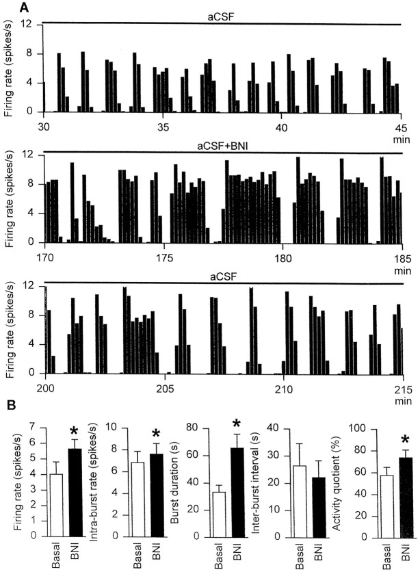
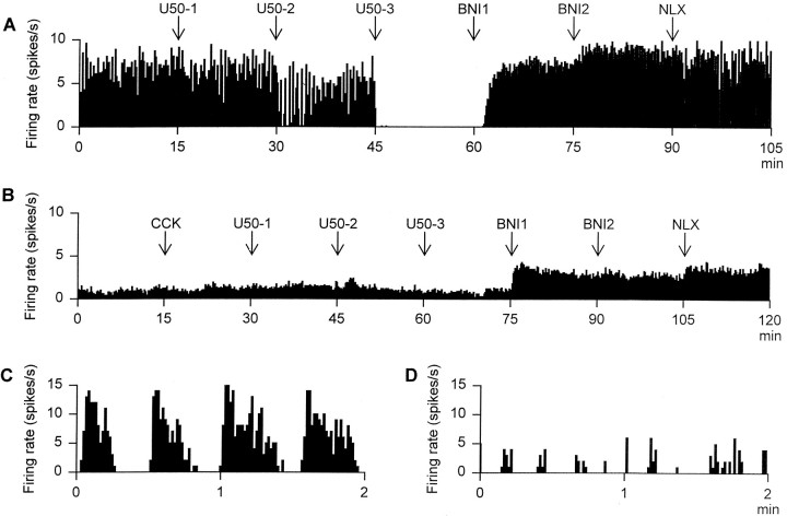
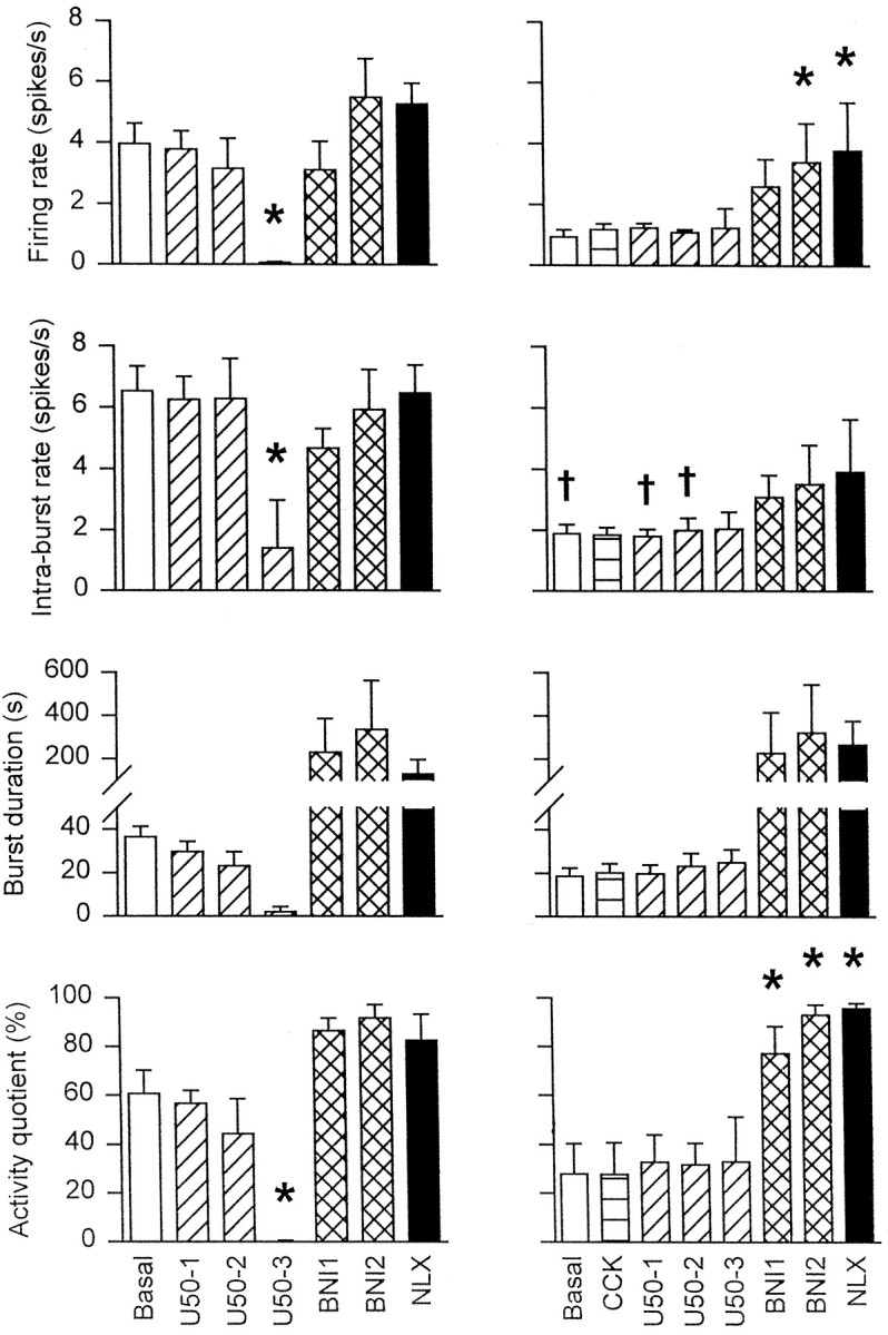
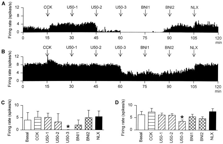
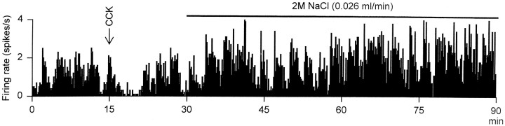
We investigated the influence of endogenous kappa-opioids on the activity of supraoptic neurons in vivo. Administration of the kappa-antagonist nor-binaltorphimine (200 micrograms/kg, i.v.), increased the activity of phasic (vasopressin), but not continuously active (oxytocin), supraoptic neurons by increasing burst duration (by 69 +/- 24%) and decreasing the interburst interval (by 19 +/- 11%). Similarly, retrodialysis of nor-binaltorphimine onto the supraoptic nucleus increased the burst duration (119 +/- 57% increase) of vasopressin cells but did not alter the firing rate of oxytocin cells (4 +/- 8% decrease). Thus, an endogenous kappa-agonist modulates vasopressin cell activity by an action within the supraoptic nucleus. To eliminate kappa-agonist actions within the supraoptic nucleus, we infused the kappa-agonist U50,488H (2.5 micrograms/hr at 0.5 micrograms/hr) into one supraoptic nucleus over 5 d to locally downregulate kappa-receptor function. Such infusions reduced the spontaneous activity of vasopressin but not oxytocin cells and reduced the proportion of cells displaying spontaneous phasic activity from 26% in vehicle-infused nuclei to 3% in U50, 488H-infused nuclei; this treatment also prevented acute inhibition of both vasopressin and oxytocin cells by U50,488H (1000 micrograms/kg, i.v.), confirming functional kappa-receptor downregulation. In U50, 488H-infused supraoptic nuclei, vasopressin cell firing rate was increased by nor-binaltorphimine (100 and 200 micrograms/kg, i.v.) but not to beyond that found in vehicle-treated nuclei, indicating that these cells were not U50,488H-dependent. Thus, normally functioning kappa-opioid mechanisms on vasopressin cells are essential for the expression of phasic firing.


Figures

Fig. 1.
Fig. 1.
A, The firing rate (averaged in 10 sec bins) of a phasic supraoptic neuron immediately before (top panel), during the final 15 min of (middle panel), and after (bottom panel) inclusion of nor-binaltorphimine (200 μg/ml) in the dialysate (aCSF at 2 μl/min) retrodialyzed directly onto the nucleus.B, The mean firing rate, intraburst firing rate, burst duration, interburst interval, and activity quotient of six phasic supraoptic nucleus neurons recorded from five rats treated as inA. *p < 0.05 versus basal; Wilcoxon signed rank test.
Fig. 2.
Fig. 2.
A, The firing rate (averaged in 10 sec bins) of a phasic cell recorded from a rat in which Ringer’s solution (0.5 μl/hr) had been infused into the supraoptic nucleus for 5 d before recording. The rat was administered the following drugs at 15 min intervals: 10 (U50-1), 100 (U50-2), and 1000 (U50-3) μg/kg intravenous U50,488H followed by 100 (BNI1) and 200 (BNI2) μg/kg intravenous nor-binaltorphimine and then 5000 μg/kg intravenous naloxone (NLX).B, The firing rate (averaged in 10 sec bins) of a continuously active vasopressin cell recorded from a rat in which U50,488H (2.5 μg/hr) had been infused into the supraoptic nucleus for 5 d before recording. The rat was administered drugs as inA, except that CCK (20 μg/kg, i.v.) was administered before the first dose of U50,488H. C, The spontaneous firing rate (in 1 sec bins) of the cell recorded inA, showing clear phasic activity. D, The spontaneous firing rate (in 1 sec bins) of an irregularly firing cell recorded from a supraoptic nucleus that had been infused with U50,488H (2.5 μg/hr) over 5 d. The cell shows “clusters” of action potentials that were typical of irregularly firing cells in U50,488H-infused rats.
Fig. 3.
Fig. 3.
The mean firing rate, intraburst firing rate, burst duration, and activity quotient of five phasic cells in Ringer’s-infused supraoptic nuclei (left panels), and five cells in U50,488H-infused supraoptic nuclei (right panels) over 15 min periods before and after administration of CCK (20 μg/kg, i.v.), U50,488H (10, 100, and 1000 μg/kg, i.v.;U50-1,U50-2, andU50-3, respectively),nor-binaltorphimine (100 and 200 μg/kg, i.v.;BNI1 and BNI2, respectively), and naloxone (5000 μg/kg, i.v.; NLX) as shown in Figure 2. Two-way RM ANOVA followed by Student Newman–Keuls test: *p < 0.05 versus basal,p < 0.05 versus matched treatment group in Ringer’s-infused rats.
Fig. 4.
Fig. 4.
The firing rate (averaged in 10 sec bins) of oxytocin cells recorded from rats in which (A) Ringer’s solution (0.5 μl/hr) or (B) U50,488H (2.5 μg/hr) had been infused into the supraoptic nucleus for 5 d before recording. The rats were intravenously administered the following drugs at 15 min intervals: 20 μg/kg CCK, 10 (U50-1), 100 (U50-2), and 1000 (U50-3) μg/kg U50,488H followed by 100 (BNI1) and 200 (BNI2) μg/kgnor-binaltorphimine, and finally 5000 μg/kg naloxone (NLX). C, The mean firing rate of oxytocin cells recorded from two control rats. D, The mean firing rate of four oxytocin cells recorded from U50,488H-infused rats. *p < 0.05 versus basal; two-way RM ANOVA followed by Student Newman–Keuls test.
Fig. 5.
Fig. 5.
The firing rate (averaged in 10 sec bins) of an irregularly active cell recorded from a rat in which U50,488H (2.5 μg/hr) had been infused into the supraoptic nucleus for 5 d before recording. The cell was inhibited by CCK (20 μg/kg, i.v.), identifying it as a vasopressin cell, and excited by intravenous infusion of 2 m NaCl (0.026 ml/min), clearly showing that this cell was not induced to fire in the robust phasic pattern typical of vasopressin cells during stimulation with hypertonic saline.

Similar articles

Cited by

References

    1. Armstrong WE, Scholer J, McNeill TH. Immunocytochemical, Golgi and electron microscopic characterization of putative dendrites in the ventral glial lamina of the rat supraoptic nucleus. Neuroscience. 1982;7:679–694. - PubMed
    1. Bicknell RJ, Chapman C, Leng G. Effects of opioid agonists and antagonists on oxytocin and vasopressin release in vitro. Neuroendocrinology. 1985;41:142–148. - PubMed
    1. Bicknell RJ, Leng G, Lincoln DW, Russell JA. Naloxone excites oxytocin neurones in the supraoptic nucleus of lactating rats after chronic morphine treatment. J Physiol (Lond) 1988;396:297–317. - PMC - PubMed
    1. Bondy CA, Gainer H, Russell JT. Dynorphin A inhibits and naloxone increases the electrically stimulated release of oxytocin but not vasopressin from the terminals of the neural lobe. Endocrinology. 1988;122:1321–1327. - PubMed
    1. Bourque CW. Intraterminal recordings from the rat neurohypophysis in vitro. J Physiol (Lond) 1990;421:247–262. - PMC - PubMed

Publication types

MeSH terms

LinkOut - more resources