. 1998 Nov;9(11):3179-93.
doi: 10.1091/mbc.9.11.3179.

Control of cyclin D1, p27(Kip1), and cell cycle progression in human capillary endothelial cells by cell shape and cytoskeletal tension

Affiliations
Free PMC article

Control of cyclin D1, p27(Kip1), and cell cycle progression in human capillary endothelial cells by cell shape and cytoskeletal tension

S Huang et al. Mol Biol Cell. 1998 Nov.
Free PMC article

Abstract

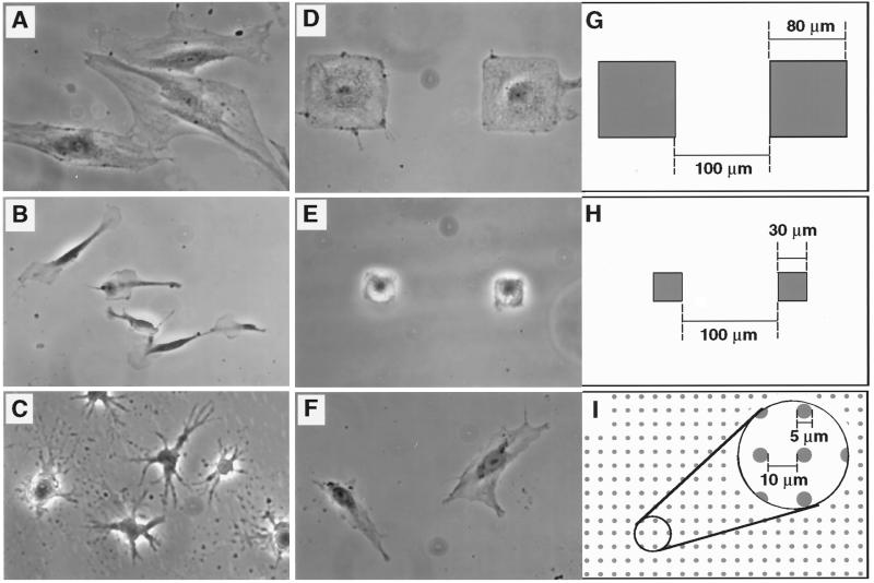
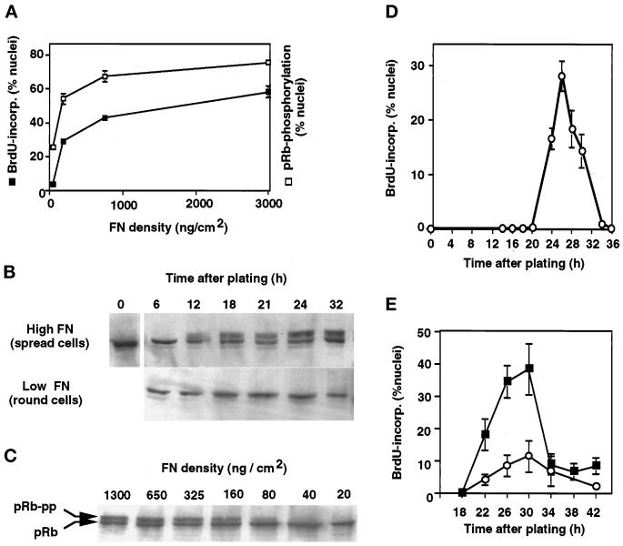
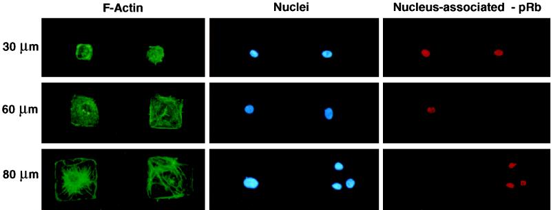
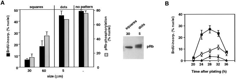
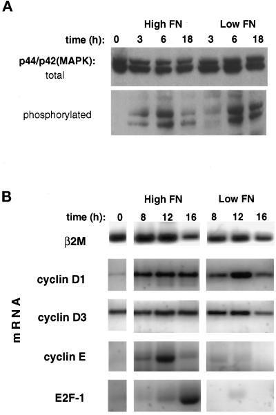
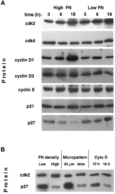
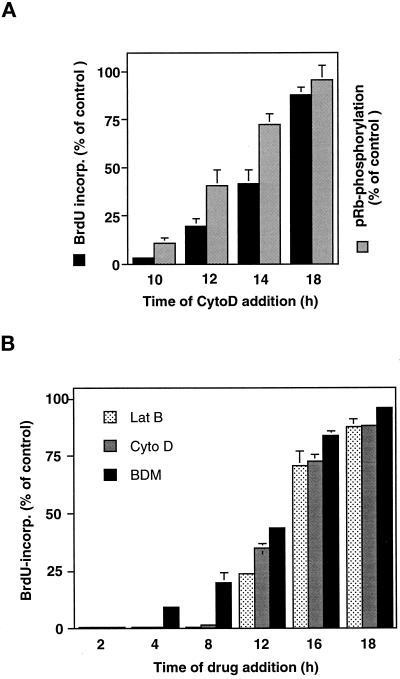
The extracellular matrix (ECM) plays an essential role in the regulation of cell proliferation during angiogenesis. Cell adhesion to ECM is mediated by binding of cell surface integrin receptors, which both activate intracellular signaling cascades and mediate tension-dependent changes in cell shape and cytoskeletal structure. Although the growth control field has focused on early integrin and growth factor signaling events, recent studies suggest that cell shape may play an equally critical role in control of cell cycle progression. Studies were carried out to determine when cell shape exerts its regulatory effects during the cell cycle and to analyze the molecular basis for shape-dependent growth control. The shape of human capillary endothelial cells was controlled by culturing cells on microfabricated substrates containing ECM-coated adhesive islands with defined shape and size on the micrometer scale or on plastic dishes coated with defined ECM molecular coating densities. Cells that were prevented from spreading in medium containing soluble growth factors exhibited normal activation of the mitogen-activated kinase (erk1/erk2) growth signaling pathway. However, in contrast to spread cells, these cells failed to progress through G1 and enter S phase. This shape-dependent block in cell cycle progression correlated with a failure to increase cyclin D1 protein levels, down-regulate the cell cycle inhibitor p27(Kip1), and phosphorylate the retinoblastoma protein in late G1. A similar block in cell cycle progression was induced before this same shape-sensitive restriction point by disrupting the actin network using cytochalasin or by inhibiting cytoskeletal tension generation using an inhibitor of actomyosin interactions. In contrast, neither modifications of cell shape, cytoskeletal structure, nor mechanical tension had any effect on S phase entry when added at later times. These findings demonstrate that although early growth factor and integrin signaling events are required for growth, they alone are not sufficient. Subsequent cell cycle progression and, hence, cell proliferation are controlled by tension-dependent changes in cell shape and cytoskeletal structure that act by subjugating the molecular machinery that regulates the G1/S transition.


Figures

Figure 1
Figure 1
Modulation of CE cell shape. (A–F) Phase contrast views of CE cells cultured on FN-coated substrates; (G–I) schematic diagrams illustrating designs used to create micropatterned adhesive islands used for studies shown in D–F, respectively (all at same magnification). (A) High-density FN (3 μg/cm2); (B) low-density FN (25 ng/cm2); (C) high-density FN in the presence of cyto D (1 μg/ml); (D and G) 80-μm-square adhesive islands; (E and H) 30-μm-square adhesive islands; (F and I) 5-μm circular adhesive islands. Note that CE cells extended over multiple small dotlike adhesive islands separated by 10-μm-wide uncoated regions, whereas they remained restricted to single larger-size islands that are separated by larger nonadhesive boundary regions (100 μm wide). Shaded areas in diagrams represent adhesive regions coated with high-density FN.
Figure 2
Figure 2
Control of G1 progression by varying cell shape through FN density. (A) Effects of varying FN coating density on DNA synthesis (cumulative BrdU incorporation into nuclei) and pRb hyperphosphorylation as measured in situ by nuclear extractability of hyperphosphorylated pRb (see MATERIALS AND METHODS). Error bars represent SD. (B) Western blots showing the time course of pRb hyperphosphorylation in spread cells on high FN compared with round cells on low FN. Cells were released from the lovastatin block at the time of plating; the slower-migrating band represents the hyperphosphorylated form of pRb. (C) Western blot demonstrating that decreasing the FN coating density inhibits pRb hyperphosphorylation (pRb-pp) when measured 18 h after release of lovastatin arrest in CE cells. (D) Kinetics of entry into S phase in lovastatin-synchronized cells on high FN as measured by pulse labeling with BrdU. (E) Kinetics of entry into S phase was similar in highly extended cells on high FN (3 μg/cm2; ▪) and in moderately spread cells on an intermediate FN density (100 ng/cm2; ○); highly retracted cells on low FN did not enter S phase.
Figure 3
Figure 3
Control of cell shape, cytoskeletal organization, nuclear size, and pRB hyperphosphorylation on micropatterned adhesive islands. Shown are fluorescence views of CE cells cultured on square adhesive islands 30, 60, or 80 μm wide and stained for F-actin using FITC-phalloidin, nuclei using the DNA binding dye DAPI, or nuclear-associated (hypophosphorylated) pRb using a monoclonal antibody. A rare occurrence can be seen at the bottom right, in which three cells are adherent to a single 80-μm-wide island; note that these three smaller cells retain the hypophosphorylated form of pRB in their nuclei, whereas the single cell that is fully spread on the adjacent island at the left lacks nuclear pRB staining and hence, underwent pRb hyperphosphorylation.
Figure 4
Figure 4
Cell shape–dependent control of passage through the G1/S transition. (A) Left, effects of varying FN substrate geometry on S phase entry (cumulative nuclear incorporation of BrdU at 30 h; black bars) and pRb hyperphosphorylation (negative pRb staining nuclei after treatment with nuclear extraction buffer at 18 h; gray bars) in synchronized CE cells, determined as in Figure 2A. Size indicates width of square islands and diameter of circular dotlike islands; no pattern indicates an unpatterned surface of identical chemistry. Right, for the 30-μm squares and the 5-μm dots, pRb phosphorylation status was confirmed by Western blot analysis as in Figure 2. (B) Kinetics of S phase entry in synchronized CE cells on unpatterned substrata (▪) or circular adhesive islands with diameters of 50 (○) or 20 μm (▴), measured as in Figure 2D.
Figure 5
Figure 5
Mapping the shape-sensitive restriction point. (A) Western blots of total cell lysates from synchronized CE cells plated on high vs. low FN probed with antibodies against total p44/p42MAPK and phosphorylated (activated) p44/p42MAPK. (B) Steady-state mRNA levels of the G1-associated genes, E2F-1, and cyclins D1, D3, and E compared with β2-microglobulin (β2 M) in CE cells grown for the indicated times on high vs. low FN, as determined using RT-PCR.
Figure 6
Figure 6
Effects of varying FN density and cell spreading on cell cycle–associated proteins. (A) Western blots of total cell lysates from cells cultured for indicated times on high vs. low FN probed with antibodies against cdks 2 and 4 and cyclins D1, D3, and E, p21Cip1(p21), or p27Kip1 (p27). (B) Western blot comparing levels of cdk2 and p27Kip1 (p27) in total cell lysates of synchronized CE cells cultured on low FN, high FN, micropatterned squares 30 μm wide (Figure 1, E and H), micropatterned dots 5 μm in diameter (Figure 1 F and I), or on high FN in the presence of cyto D (1 μg/ml) added at the indicated times. All lysates were obtained 18 h after plating.
Figure 7
Figure 7
Role of the actin cytoskeleton and cytoskeletal tension in G1 progression. (A) Cyto D (1 μg/ml) was added at the indicated times to cultures of synchronized CE cells plated on high FN. Cumulative BrdU incorporation into DNA (black bars) and pRb hyperphosphorylation (stippled bars) were measured at 30 and 20 h, respectively, as described in Figure 2, and shown as relative values compared with the untreated control cultures. Error bars indicate SD. (B) Lat B (0.1 μg/ml), cyto D (1 μg/ml), and BDM (20 mM), as indicated in the inset legend, were added at the indicated times and BrdU incorporation determined as in A in parallel studies.

References

    1. Alessandrini A, Chiaur DS, Pagano M. Regulation of the cyclin-dependent kinase inhibitor p27 by degradation and phosphorylation. Leukemia. 1997;11:342–345. - PubMed
    1. Anderson NG, Maller JL, Tonks NK, Sturgill TW. Requirement for integration of signals from two distinct phosphorylation pathways for activation of MAP kinase. Nature. 1990;343:651–653. - PubMed
    1. Assoian RK, Zhu X. Cell anchorage and the cytoskeleton as partners in growth factor dependent cell cycle progression. Curr Opin Cell Biol. 1997;9:93–98. - PubMed
    1. Baldin V, Lukas J, Marcote MJ, Pagano M, Draetta G. Cyclin D1 is a nuclear protein required for cell cycle progression in G1. Genes Dev. 1993;7:812–821. - PubMed
    1. Ben Ze’ev A, Farmer SR, Penman S. Protein synthesis requires cell-surface contact while nuclear events respond to cell shape in anchorage-dependent fibroblasts. Cell. 1980;21:365–372. - PubMed

Publication types