. 1998 Nov;9(11):3241-57.
doi: 10.1091/mbc.9.11.3241.

Rab11 is required for trans-golgi network-to-plasma membrane transport and a preferential target for GDP dissociation inhibitor

Affiliations
Free PMC article

Rab11 is required for trans-golgi network-to-plasma membrane transport and a preferential target for GDP dissociation inhibitor

W Chen et al. Mol Biol Cell. 1998 Nov.
Free PMC article

Abstract

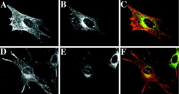
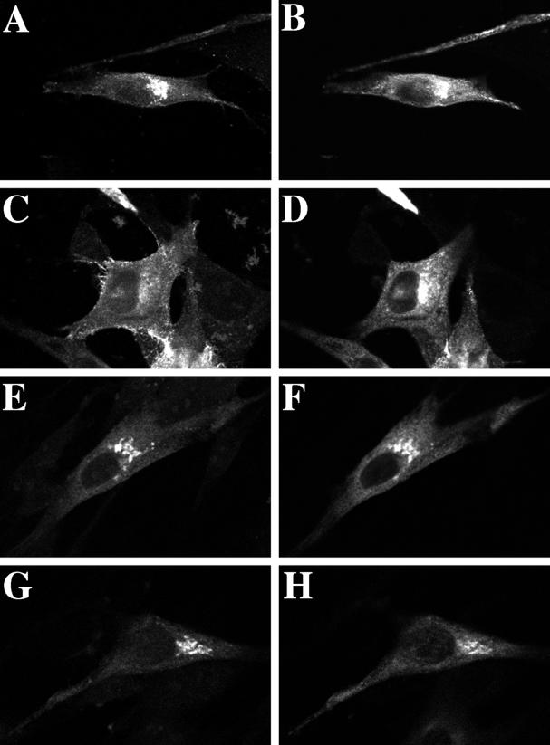
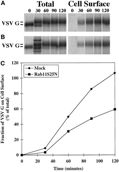
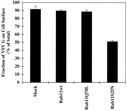
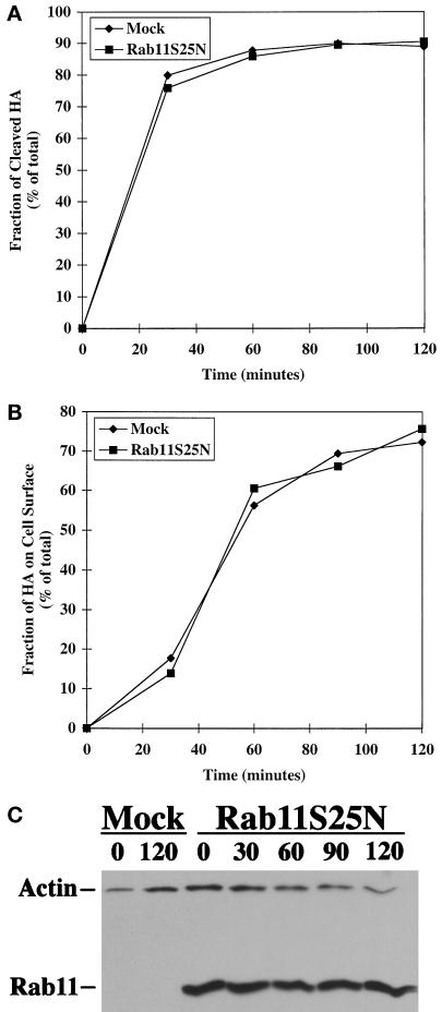
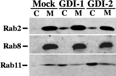
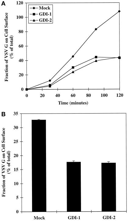
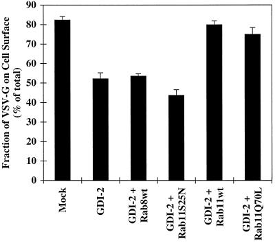
The rab11 GTPase has been localized to both the Golgi and recycling endosomes; however, its Golgi-associated function has remained obscure. In this study, rab11 function in exocytic transport was analyzed by using two independent means to perturb its activity. First, expression of the dominant interfering rab11S25N mutant protein led to a significant inhibition of the cell surface transport of vesicular stomatitis virus (VSV) G protein and caused VSV G protein to accumulate in the Golgi. On the other hand, the expression of wild-type rab11 or the activating rab11Q70L mutant had no adverse effect on VSV G transport. Next, the membrane association of rab11, which is crucial for its function, was perturbed by modest increases in GDP dissociation inhibitor (GDI) levels. This led to selective inhibition of the trans-Golgi network to cell surface delivery, whereas endoplasmic reticulum-to-Golgi and intra-Golgi transport were largely unaffected. The transport inhibition was reversed specifically by coexpression of wild-type rab11 with GDI. Under the same conditions two other exocytic rab proteins, rab2 and rab8, remained membrane bound, and the transport steps regulated by these rab proteins were unaffected. Neither mutant rab11S25N nor GDI overexpression had any impact on the cell surface delivery of influenza hemagglutinin. These data show that functional rab11 is critical for the export of a basolateral marker but not an apical marker from the trans-Golgi network and pinpoint rab11 as a sensitive target for inhibition by excess GDI.


Figures

Figure 1
Figure 1
Localization of rab11 and the Golgi marker α2,6-ST in BHK cells. Myc epitope-tagged ST was transiently overexpressed in BHK cells under the control of a constitutive CMV promoter together with GFP-tagged wild-type rab11 (A–C) or rab11S25N (D–F). At 17 h after transfection, cells were incubated at 20°C for 2 h to increase the ST signal in the Golgi (Ma et al., 1997). Cells were then fixed and processed for confocal microscopy. GFP-tagged rab11 proteins were visualized directly (A and D), whereas ST was detected using a mouse anti-myc antibody as the primary antibody and a horse anti-mouse, Texas Red–conjugated secondary antibody (B and E). Each image represents a single 0.4-μm section with ST and GFP-tagged rab11 proteins viewed in the same focal plane. (C and F) Merged images.
Figure 2
Figure 2
Localization of rab11 and transferrin receptor in BHK cells. BHK cells were cotransfected with human transferrin receptor and GFP-tagged wild-type rab11 (A–C) or rab11S25N (D–F). At 17 h after transfection, cells were fixed and processed for confocal microscopy. Transferrin receptor was visualized using a goat anti-transferrin receptor antibody as the primary antibody and a biotinylated horse anti-goat antibody and Texas Red–conjugated avidin for detection purposes (A and D). GFP-tagged rab11 proteins were visualized directly (B and E). Each image represents a single 0.4-μm section with transferrin receptor and GFP-tagged rab11 proteins viewed in the same focal plane. (C and F) Merged images. The arrows in B designate some of the peripheral endosomes positive for wild-type rab11 and transferrin receptor.
Figure 3
Figure 3
Overexpression of the dominant negative rab11S25N mutant causes VSV G protein to accumulate in the Golgi. VSV G protein was transiently overexpressed in BHK cells under the control of a constitutive CMV promoter in conjunction with GFP-tagged wild-type rab11 (A–D) or rab11S25N (E–H). At 17 h after transfection, cells were incubated at 20°C for 2 h (A, B, E, and F). Parallel samples were transferred to 37°C for 1 h immediately after the 20°C incubation (C, D, G, and H). Cells were then fixed and processed for confocal microscopy. VSV G protein was detected using mouse mAb P5D4 as the primary antibody and a horse anti-mouse, Texas Red–conjugated secondary antibody (A, C, E, and G). GFP-tagged rab11 proteins were visualized directly (B, D, F, and H). Each image represents a single 0.4-μm section with VSV G protein (left panels) and GFP-tagged rab11 proteins (right panels) viewed in the same focal plane.
Figure 4
Figure 4
The kinetics of VSV G protein cell surface delivery is markedly altered by overexpression of the dominant negative rab11S25N mutant. Rab11S25N was coexpressed with VSV G protein in BHK cells. At 5 h after transfection, cells were metabolically labeled for 10 min at 37°C and subsequently incubated for various chase times (0–120 min). The kinetics of cell surface delivery was monitored by biotinylation at 4°C. Biotinylated (cell surface) and total cellular VSV G fractions were prepared, analyzed by SDS-PAGE, and quantified with a Fuji Bioimager. Quantitation of VSV G protein cell surface appearance in mock-treated control cells (A) and cells overexpressing rab11S25N mutant (B) is shown in C. (A and B) Left panels, quantitative immunoprecipitation of total VSV G protein from cell lysates after different chase periods using mouse mAb P5D4 (Note that there are two unidentified background bands in the bottom 30-min lane); right panels, cell surface, biotinylated VSV G protein recovered using streptavidin-Sepharose after different chase periods. Representative data from one of three independent trials are shown.
Figure 5
Figure 5
Cell surface delivery of VSV G protein is reduced in cells expressing the dominant negative rab11S25N mutant. BHK cells were transfected with VSV G together with wild-type (wt) rab11, rab11Q70L, or rab11S25N. At 5 h after transfection, cells were metabolically labeled for 10 min. After a chase period of 120 min, cell surface delivery of the VSV G protein was monitored by surface biotinylation as described in MATERIALS AND METHODS. Each column represents the mean ± SEM of triplicate samples from one of three independent experiments.
Figure 6
Figure 6
Exocytosis of influenza HA is unaffected by overexpression of the dominant negative rab11S25N mutant. Rab11S25N was coexpressed with influenza HA in BHK cells. At 5 h after transfection, cells were metabolically labeled for 10 min at 37°C and subsequently incubated for various chase times (0–120 min). Cell surface biotinylation and immunoprecipitation were performed as described in MATERIALS AND METHODS. (A) Quantitation of the kinetics of FPV HA cleavage as a measure of transport through the TGN. (B) Quantitation of influenza HA cell surface appearance in cells overexpressing rab11S25N compared with mock-treated control cells. (C) Detection of rab11 in transfected cell lysates by immunoblotting with actin serving as a control for protein loading.
Figure 7
Figure 7
Distribution of rab2, rab8, and rab11 between cytosol and total membrane fractions after GDI overexpression. VSV G epitope-tagged GDI-1 or GDI-2 proteins were overexpressed in BHK cells; 6 h after transfection, the cells were homogenized, and cytosol and total membrane fractions were prepared. Cytosol (C) and total membrane (M) fractions were resolved by SDS-PAGE. The proteins were transferred to polyvinylidene difluoride membranes, and blots were probed individually with antibodies against rab2, rab8, or rab11. An HRP-conjugated secondary antibody was used to detect the immune complexes by chemiluminescence.
Figure 8
Figure 8
TGN–to–cell surface transport of VSV G tsO45 protein is inhibited by overexpression of either GDI isoform. GDI-1 and GDI-2 tagged with a VSV G epitope were coexpressed with the temperature-sensitive VSV G tsO45 protein in BHK cells. This enabled the simultaneous monitoring of both overexpressed proteins. Transfected cells were maintained and metabolically labeled (10 min, 5 h after transfection) at nonpermissive temperature (39°C). Cell surface transport of VSV G tsO45 protein was induced by transfer to permissive temperature (31°C) for various chase times (0–120 min). (A) The cell surface delivery of the VSV G protein as a function of time was monitored by surface biotinylation as described in MATERIALS AND METHODS. Representative data from one of three independent experiments is shown. (B) TGN–to–cell surface transport of VSV G tsO45 proteins was monitored by transferring transfected cells from 39 to 20°C for 1 h to accumulate VSV G in the TGN and subsequently transferring them to 31°C for 1 h. Cell surface delivery was quantified by biotinylation as described in MATERIALS AND METHODS. Each column represents the mean ± SEM of triplicate samples from one of two independent experiments.
Figure 9
Figure 9
The inhibition of cell surface delivery of VSV G tsO45 protein by GDI-2 overexpression can be relieved by coexpression of wild-type rab11 but not wild-type rab8. BHK cells were transfected with the VSV G tsO45 plasmid (0.3 μg/35-mm dish) and GDI-2 epitope tagged with VSV G plasmid (0.6 μg/35-mm dish) together with wild-type (wt) rab8, rab11S25N, wild-type (wt) rab11, or rab11Q70L (0.3 μg/35-mm dish). At 5 h after transfection, cells were metabolically labeled for 10 min and then subjected to a 90-min chase period. The cell surface delivery of the VSV G tsO45 protein was monitored by surface biotinylation as described in MATERIALS AND METHODS. Each column represents the mean ± SEM of triplicate samples from one of three independent experiments.

References

    1. Bachner D, Sedlacek Z, Korn B, Hameister H, Poustka A. Expression patterns of two human genes coding for different rab GDP-dissociation inhibitors (GDIs), extremely conserved proteins involved in cellular transport. Hum Mol Genet. 1995;4:701–708. - PubMed
    1. Benli M, Döring F, Robinson DG, Yang X, Gallwitz D. Two GTPase isoforms, Ypt31p and Ypt32p, are essential for Golgi function in yeast. EMBO J. 1996;15:6460–6475. - PMC - PubMed
    1. Brent R, Ptashne M. A eukaryotic transcriptional activator bearing the DNA specificity of a prokaryotic repressor. Cell. 1985;43:729–736. - PubMed
    1. Bucci C, Parton RG, Mather IH, Stunnenberg H, Simons K, Hoflack B, Zerial M. The small GTPase rab5 functions as a regulatory factor in the early endocytic pathway. Cell. 1992;70:715–728. - PubMed
    1. Calhoun BC, Goldenring JR. Rab proteins in gastric parietal cells: evidence for the membrane recycling hypothesis. Yale J Biol Med. 1997;69:1–8. - PMC - PubMed

Publication types

Substances

Associated data

LinkOut - more resources