. 1998 Nov 2;188(9):1641-50.
doi: 10.1084/jem.188.9.1641.

Ex vivo staining of metastatic lymph nodes by class I major histocompatibility complex tetramers reveals high numbers of antigen-experienced tumor-specific cytolytic T lymphocytes

Affiliations

Ex vivo staining of metastatic lymph nodes by class I major histocompatibility complex tetramers reveals high numbers of antigen-experienced tumor-specific cytolytic T lymphocytes

P Romero et al. J Exp Med. .

Abstract

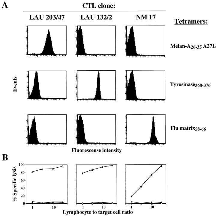
Characterization of cytolytic T lymphocyte (CTL) responses to tumor antigens has been impeded by a lack of direct assays of CTL activity. We have synthesized reagents ("tetramers") that specifically stain CTLs recognizing melanoma antigens. Tetramer staining of tumor-infiltrated lymph nodes ex vivo revealed high frequencies of tumor-specific CTLs which were antigen-experienced by surface phenotype. In vitro culture of lymph node cells with cytokines resulted in very large expansions of tumor-specific CTLs that were dependent on the presence of tumor cells in the lymph nodes. Tetramer-guided sorting by flow cytometer allowed isolation of melanoma-specific CTLs and confirmation of their specificity and their ability to lyse autologous tumor cells. Our results demonstrate the value of these novel reagents for monitoring tumor-specific CTL responses and for generating CTLs for adoptive immunotherapy. These data also indicate that strong CTL responses to melanoma often occur in vivo, and that the reactive CTLs have substantial proliferative and tumoricidal potential.


Figures

Figure 2
Figure 2
Direct identification of HLA-A2/tumor peptide antigen tetramer binding lymphocytes in TILNs. (A) Four TILNs obtained from three HLA-A2 melanoma patients were stained, after overnight culture, with either A2/Melan-A26–35 A27L or A2/tyrosinase368–376 tetramers together with anti-CD3PerCP mAb and anti-CD8FITC mAb. Dot plots are shown for gated CD8+ LN cells. (B) Ex vivo A2/Melan-A tetramer+ TILNs have an activated phenotype. Cell suspensions prepared from normal (NLN) or metastatic (TILN) LNs from patient LAU 267 were directly analyzed by three-color flow cytometry using anti-CD8PerCP mAb, A2/Melan-A tetramers, and either anti-CD45RAFITC mAb (top) or anti-CD45ROFITC mAb (bottom). Dot plots are shown for gated CD8+ LN cells. (C) Enumeration and phenotype of tetramer+ cells in ex vivo circulating lymphocytes. Highly homogeneous CD8+ lymphocyte populations (>98%) were obtained from PBMC of patient LAU 267, or from a healthy donor (HD), by two rounds of positive selection with magnetic cell sorting. The lymphocyte preparation was then stained with A2/Melan-A or A2/influenza matrix tetramers and anti-CD45RACychrome and analyzed immediately by flow cytometry. The histograms on top of dot plots show the intensity of the fluorescence signal on the FL-3 channel of gated Melan-A/tetramer+ or influenza tetramer+ lymphocytes. (D) Summary of phenotyping data obtained from NLNs, TILNs, and PBMC from patients LAU 181 and LAU 267. The LN cell suspensions were stained either immediately after cell suspension preparation (LAU 267) or after overnight culture of previously cryopreserved uncultured LN cells (LAU 181) with either anti-CD45RAFITC or anti-CD45ROFITC in combination with anti-CD8PerP and A2/Melan-A tetramers. The results for the CD45RA and CD45RO triple staining of NLNs and TILN-2 from LAU 267 are illustrated in B. Additionally, PBMC available from the same time of the LN dissection from patient LAU 267 were stained as illustrated in C. The percentages of either CD45RO+ or CD45RA in gated A2/Melan-A tetramer+ LN cells were calculated with CellQuest™ software.
Figure 2
Figure 2
Direct identification of HLA-A2/tumor peptide antigen tetramer binding lymphocytes in TILNs. (A) Four TILNs obtained from three HLA-A2 melanoma patients were stained, after overnight culture, with either A2/Melan-A26–35 A27L or A2/tyrosinase368–376 tetramers together with anti-CD3PerCP mAb and anti-CD8FITC mAb. Dot plots are shown for gated CD8+ LN cells. (B) Ex vivo A2/Melan-A tetramer+ TILNs have an activated phenotype. Cell suspensions prepared from normal (NLN) or metastatic (TILN) LNs from patient LAU 267 were directly analyzed by three-color flow cytometry using anti-CD8PerCP mAb, A2/Melan-A tetramers, and either anti-CD45RAFITC mAb (top) or anti-CD45ROFITC mAb (bottom). Dot plots are shown for gated CD8+ LN cells. (C) Enumeration and phenotype of tetramer+ cells in ex vivo circulating lymphocytes. Highly homogeneous CD8+ lymphocyte populations (>98%) were obtained from PBMC of patient LAU 267, or from a healthy donor (HD), by two rounds of positive selection with magnetic cell sorting. The lymphocyte preparation was then stained with A2/Melan-A or A2/influenza matrix tetramers and anti-CD45RACychrome and analyzed immediately by flow cytometry. The histograms on top of dot plots show the intensity of the fluorescence signal on the FL-3 channel of gated Melan-A/tetramer+ or influenza tetramer+ lymphocytes. (D) Summary of phenotyping data obtained from NLNs, TILNs, and PBMC from patients LAU 181 and LAU 267. The LN cell suspensions were stained either immediately after cell suspension preparation (LAU 267) or after overnight culture of previously cryopreserved uncultured LN cells (LAU 181) with either anti-CD45RAFITC or anti-CD45ROFITC in combination with anti-CD8PerP and A2/Melan-A tetramers. The results for the CD45RA and CD45RO triple staining of NLNs and TILN-2 from LAU 267 are illustrated in B. Additionally, PBMC available from the same time of the LN dissection from patient LAU 267 were stained as illustrated in C. The percentages of either CD45RO+ or CD45RA in gated A2/Melan-A tetramer+ LN cells were calculated with CellQuest™ software.
Figure 1
Figure 1
Fluorescent soluble HLA-A2/ antigenic peptide tetramers specifically stain cloned CTLs. (A) Three HLA-A2/peptide tetramers, incorporating Melan-A26–35 A27L analogue, tyrosinase368–376, and influenza matrix58–66 peptides, were tested for their ability to stain CTL clones LAU 203/47 specific for Melan-A26–35, LAU 132/2 specific for tyrosinase368–376, and NM 17 specific for influenza matrix58–66. (B) The specificity of peptide antigen recognition by the three CTL clones was assayed on chromium-labeled T2 target cells sensitized with either Melan-A26–35 (open triangles), tyrosinase368–376 (filled triangles), or influenza matrix58–66 (filled circles) at the indicated lymphocyte to target cell ratios.
Figure 3
Figure 3
Functional activity of TILNs sorted according to their tetramer staining phenotype correlates with antigen specificity. (A) Cell suspensions prepared from a metastatic (TILN) and a normal (NLN) LN excised from the same anatomical region of patient LAU 181 were cultured in complete medium supplemented with rIL-2 and rIL-7, and aliquots of cells were triple stained with anti-CD8FITC mAb, anti-CD3PerCP mAb, and A2/Melan-A tetramers either after overnight (top), 15 d (middle), or 21 d (bottom) of culture. A2/Melan-A tetramer versus CD3 dot plots are shown for gated CD8+ live lymphocytes, and the percentage of cells staining positively with the tetramers is indicated. (B) The TILN population from patient LAU 233 was analyzed by three-color cytometry, after 12 d in culture with recombinant cytokines only, using the same reagents as in A, and the staining profiles are shown (top, Unsorted TILN). CD3+ CD8+ TILNs were sterile sorted into A2/ Melan-A tetramer+ and tetramer populations. After expansion for 2 wk in the presence of irradiated allogeneic PBMC and PHA, the sorted cells were stained as above, and results are shown for the tetramer cell fraction (middle) and the tetramer+ cell fraction (bottom). (C) Each cell fraction was tested for its lytic activity against chromium-labeled T2 cells which were sensitized separately with the following HLA-A2– restricted melanoma-associated peptides: T2 cells alone (open circles, broken lines), T2 + gp100 154– 162 (filled circles, broken lines), T2 + gp100 209– 217 (open triangles, broken lines), T2 + gp100 280–288 (filled triangles, broken lines), T2 + gp100 457–466 (open squares), T2 + gp100 476–485 (filled squares), T2 + tyrosinase 1–9 (open circles), T2 + tyrosinase 368–376 (filled circles), T2 + Melan-A 26–35 (open triangles), T2 + MAGE-3 271–279 (filled triangles). (D) The same cell fractions assayed above, unsorted population (open circles), tetramer+ (filled triangles), and tetramer (open triangles), were also assayed for their lytic activity against the autologous tumor cell line, Me 305, derived from the same TILN cell population.
Figure 3
Figure 3
Functional activity of TILNs sorted according to their tetramer staining phenotype correlates with antigen specificity. (A) Cell suspensions prepared from a metastatic (TILN) and a normal (NLN) LN excised from the same anatomical region of patient LAU 181 were cultured in complete medium supplemented with rIL-2 and rIL-7, and aliquots of cells were triple stained with anti-CD8FITC mAb, anti-CD3PerCP mAb, and A2/Melan-A tetramers either after overnight (top), 15 d (middle), or 21 d (bottom) of culture. A2/Melan-A tetramer versus CD3 dot plots are shown for gated CD8+ live lymphocytes, and the percentage of cells staining positively with the tetramers is indicated. (B) The TILN population from patient LAU 233 was analyzed by three-color cytometry, after 12 d in culture with recombinant cytokines only, using the same reagents as in A, and the staining profiles are shown (top, Unsorted TILN). CD3+ CD8+ TILNs were sterile sorted into A2/ Melan-A tetramer+ and tetramer populations. After expansion for 2 wk in the presence of irradiated allogeneic PBMC and PHA, the sorted cells were stained as above, and results are shown for the tetramer cell fraction (middle) and the tetramer+ cell fraction (bottom). (C) Each cell fraction was tested for its lytic activity against chromium-labeled T2 cells which were sensitized separately with the following HLA-A2– restricted melanoma-associated peptides: T2 cells alone (open circles, broken lines), T2 + gp100 154– 162 (filled circles, broken lines), T2 + gp100 209– 217 (open triangles, broken lines), T2 + gp100 280–288 (filled triangles, broken lines), T2 + gp100 457–466 (open squares), T2 + gp100 476–485 (filled squares), T2 + tyrosinase 1–9 (open circles), T2 + tyrosinase 368–376 (filled circles), T2 + Melan-A 26–35 (open triangles), T2 + MAGE-3 271–279 (filled triangles). (D) The same cell fractions assayed above, unsorted population (open circles), tetramer+ (filled triangles), and tetramer (open triangles), were also assayed for their lytic activity against the autologous tumor cell line, Me 305, derived from the same TILN cell population.

References

    1. Robbins PF, Kawakami Y. Human tumor antigens recognized by T cells. Curr Opin Immunol. 1996;8:628–636. - PubMed
    1. Romero P. Cytolytic T lymphocyte responses of cancer patients to tumor-associated antigens. Springer Semin Immunopathol. 1996;18:185–198. - PubMed
    1. Romero P, Cerottini JC, Waanders G. Novel methods to monitor antigen-specific cytotoxic T cell responses in cancer immunotherapy. Mol Med Today. 1998;4:305–312. - PubMed
    1. Altman JD, Moss PAH, Goulder PJR, Barouch DH, McHeyzer-Williams MG, Bell JI, McMichael AJ, Davis MM. Phenotypic analysis of antigen-specific T lymphocytes. Science. 1996;274:94–96. - PubMed
    1. Dunbar PR, Ogg GS, Chen J, Rust N, van der Bruggen P, Cerundolo V. Direct isolation, phenotyping and cloning of low frequency antigen-specific cytotoxic T lymphocytes from peripheral blood. Curr Biol. 1998;8:413–416. - PubMed

Publication types

MeSH terms