. 1998 Nov 2;188(9):1657-68.
doi: 10.1084/jem.188.9.1657.

Characterization of gene expression in resting and activated mast cells

Affiliations

Characterization of gene expression in resting and activated mast cells

H Chen et al. J Exp Med. .

Erratum in

  • J Exp Med 1998 Dec 21;188(12):2387

Abstract

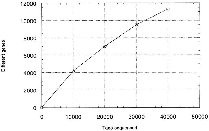
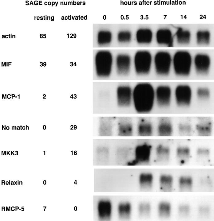
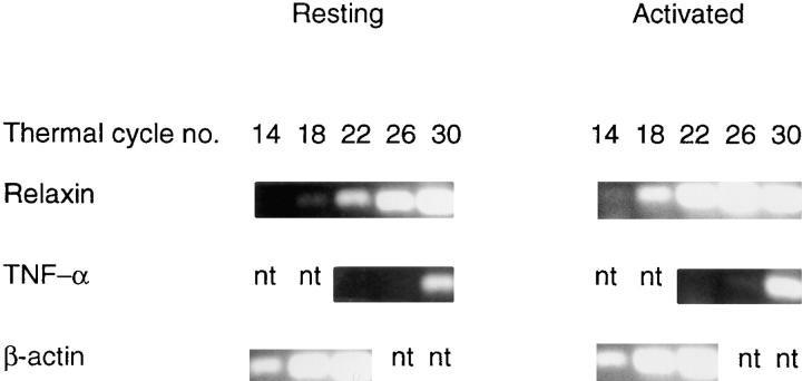
To characterize gene expression in activated mast cells more comprehensively than heretofore, we surveyed the changes in genetic transcripts by the method of serial analysis of gene expression in the RBL-2H3 line of rat mast cells before and after they were stimulated through their receptors with high affinity for immunoglobulin E (FcepsilonRI). A total of 40,759 transcripts derived from 11,300 genes were analyzed. Among the diverse genes that had not been previously associated with mast cells and that were constitutively expressed were those for the cytokine macrophage migration inhibitory factor neurohormone receptors such as growth hormone- releasing factor and melatonin and components of the exocytotic machinery. In addition, several dozen transcripts were differentially expressed in response to antigen-induced clustering of the FcepsilonRI. Included among these were the genes for preprorelaxin, mitogen-activated protein kinase kinase 3, and the dual specificity protein phosphatase, rVH6. Significantly, the majority of genes differentially expressed in this well-studied model of mast cell activation have not been identified before this analysis.


Figures

Figure 1
Figure 1
Results of the combined analysis of two SAGE tag libraries.
Figure 2
Figure 2
Characterization of transcripts and their products. (A) MIF detected by RT-PCR. Total RNAs were prepared from rat peritoneal mast cells and macrophages and used for RT-PCR using specific primers for rat MIF. The marker (leftmost lane) shows the position of a cDNA product having the expected size of 550 bp. (B) MIF detected by Western blot. Immunoprecipitates were prepared by reacting goat polyclonal anti-human MIF antibodies with the material indicated below, separated on polyacrylamide gels, and blotted either with a mouse monoclonal anti-human MIF antibody (lanes 1–6), or with polyclonal rabbit anti–rat MIF antibodies (lanes 7–9). Lanes 1, 4, and 7 were loaded with lysates from 3 × 10⁶ HMC-1, KU812, and RBL-2H3 cells, respectively; lanes 2, 5, and 8 with 1 ml of culture supernatants from HMC-1, KU812, and RBL-2H3 cells; lanes 3, 6, and 9 with 1 ml of the medium used to culture HMC-1, KU812, and RBL-2H3 cells, as a control. (C) MAPKK3 detected by Western blot. Lysates from 2 × 10⁵ RBL-2H3 cells were blotted with anti-MAPKK3 antibody. Lane 1, Unstimulated cells. Lanes 2–5, Cells stimulated for 0.5, 3.5, 7, and 16 h.
Figure 2
Figure 2
Characterization of transcripts and their products. (A) MIF detected by RT-PCR. Total RNAs were prepared from rat peritoneal mast cells and macrophages and used for RT-PCR using specific primers for rat MIF. The marker (leftmost lane) shows the position of a cDNA product having the expected size of 550 bp. (B) MIF detected by Western blot. Immunoprecipitates were prepared by reacting goat polyclonal anti-human MIF antibodies with the material indicated below, separated on polyacrylamide gels, and blotted either with a mouse monoclonal anti-human MIF antibody (lanes 1–6), or with polyclonal rabbit anti–rat MIF antibodies (lanes 7–9). Lanes 1, 4, and 7 were loaded with lysates from 3 × 10⁶ HMC-1, KU812, and RBL-2H3 cells, respectively; lanes 2, 5, and 8 with 1 ml of culture supernatants from HMC-1, KU812, and RBL-2H3 cells; lanes 3, 6, and 9 with 1 ml of the medium used to culture HMC-1, KU812, and RBL-2H3 cells, as a control. (C) MAPKK3 detected by Western blot. Lysates from 2 × 10⁵ RBL-2H3 cells were blotted with anti-MAPKK3 antibody. Lane 1, Unstimulated cells. Lanes 2–5, Cells stimulated for 0.5, 3.5, 7, and 16 h.
Figure 3
Figure 3
Northern blots of selected genes. Messenger RNAs were prepared and used for Northern blotting with end-labeled 15–40-bp oligonucleotides in all cases except for RMCP-5; for the latter a randomly labeled PCR-amplified cDNA was used. The β-actin blot is a loading control. The columns to the left of the blots list the copy numbers of the tags observed in the resting and activated SAGE libraries for the indicated genes. MKK3 refers to MAPKK3.
Figure 4
Figure 4
Results of RT-PCR amplification of mRNAs from resting and activated cells using primers specific for selected transcripts. Items shown as “nt” were not tested.

References

    1. Abraham SN, Malaviya R. Mast cells in infection and immunity. Infect Immun. 1997;65:3501–3508. - PMC - PubMed
    1. Selye, H. 1965. The Mast Cells. Butterworth Inc., Washington DC. 1–498.
    1. Rigby, L., M.D. Hulett, R.I. Brinkworth, and P.M. Hogarth. 1998. The structural basis of the interaction of IgE and FcεRI. In IgE Receptor (FcεRI) Function in Mast Cells and Basophils. M.M. Hamawy, editor. R.G. Landes Company, Austin, TX. 7–32.
    1. Hamawy, M.M., and W.D. Swaim. 1997. FcεRI-mediated cell degranulation, proliferation and adhesion. In IgE Receptor (FcεRI) Function in Mast Cells and Basophils. M.M. Hamawy, editor. R.G. Landes Company, Austin, TX. 173–180.
    1. Gordon, J.R. 1997. FcεRI-induced cytokine production and gene expression. In IgE Receptor (FcεRI) Function in Mast Cells and Basophils. M.M. Hamawy, editor. R.G. Landes Company, Austin, TX. 209–242.