. 1998 Nov 2;188(9):1691-703.
doi: 10.1084/jem.188.9.1691.

Identification of functional human splenic memory B cells by expression of CD148 and CD27

Affiliations

Identification of functional human splenic memory B cells by expression of CD148 and CD27

S G Tangye et al. J Exp Med. .

Abstract

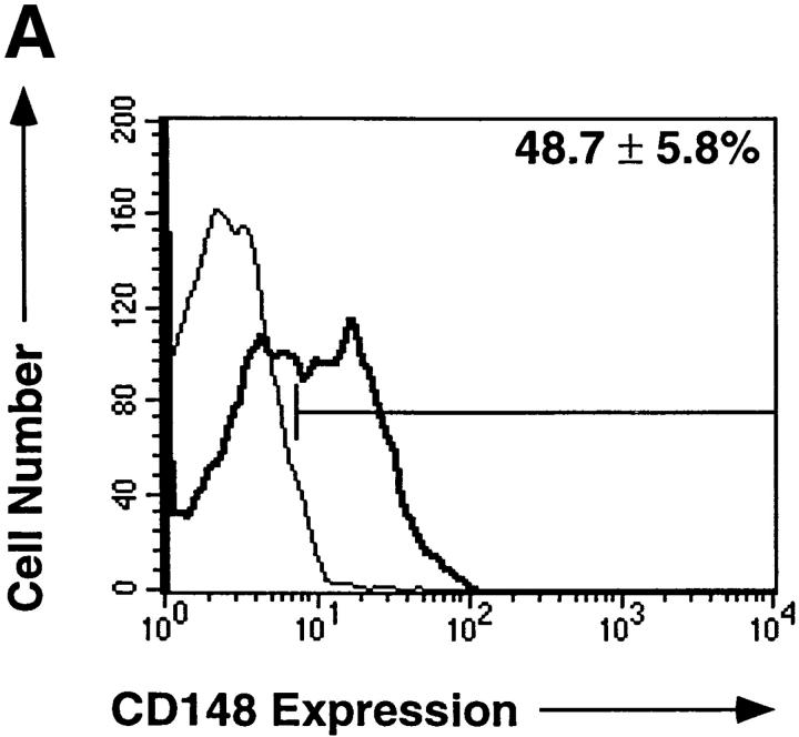
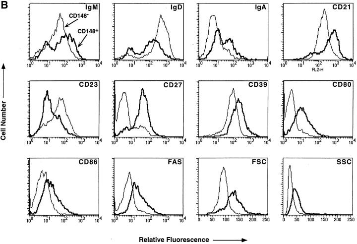
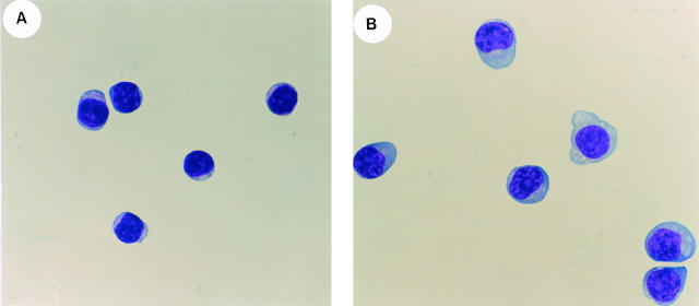
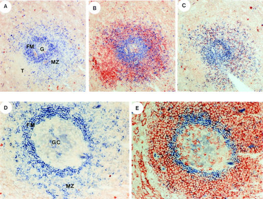
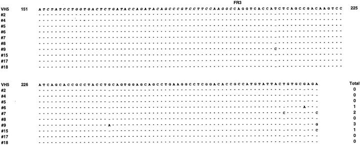
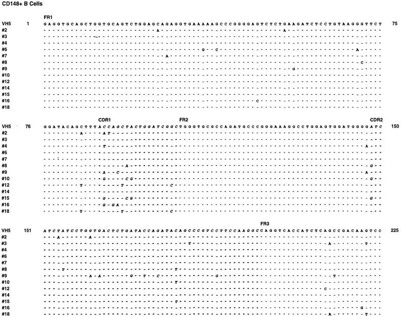
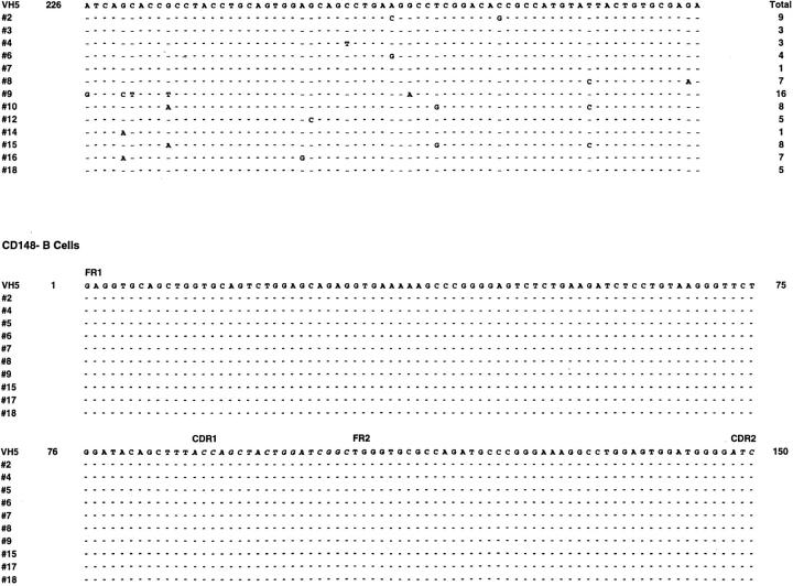
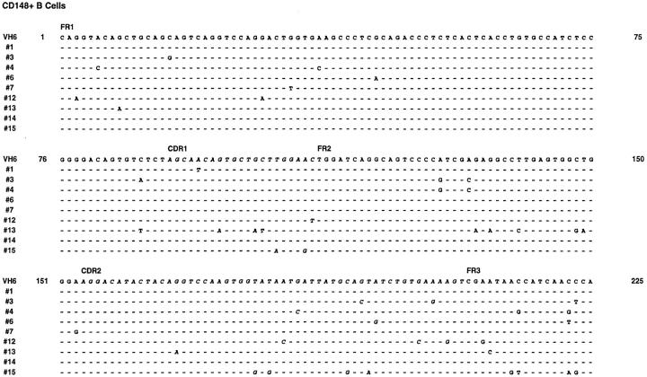
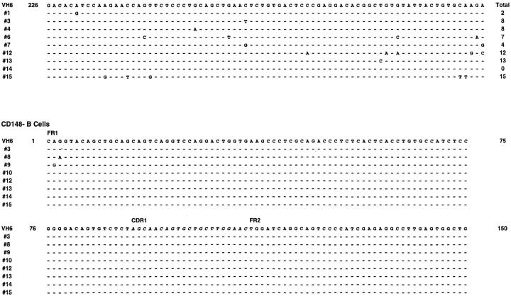
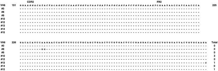
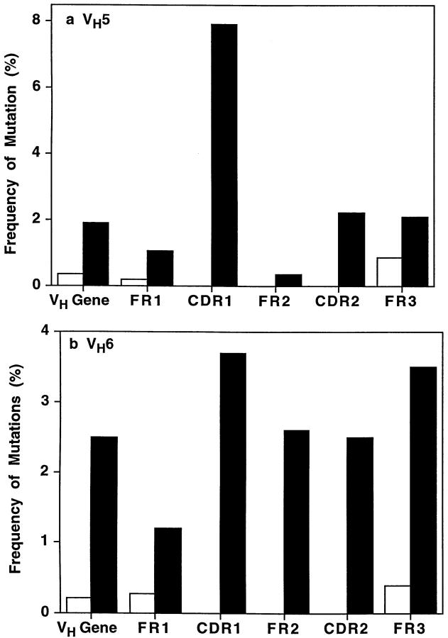
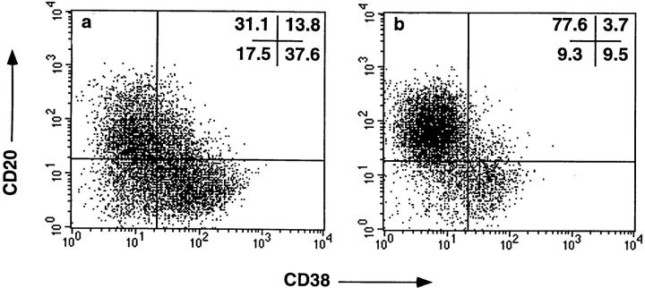
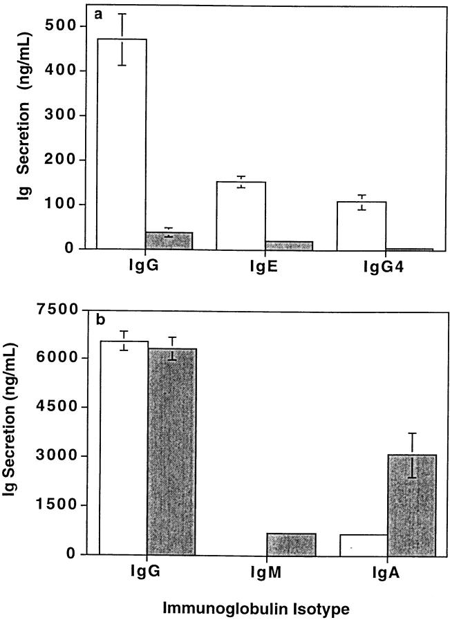
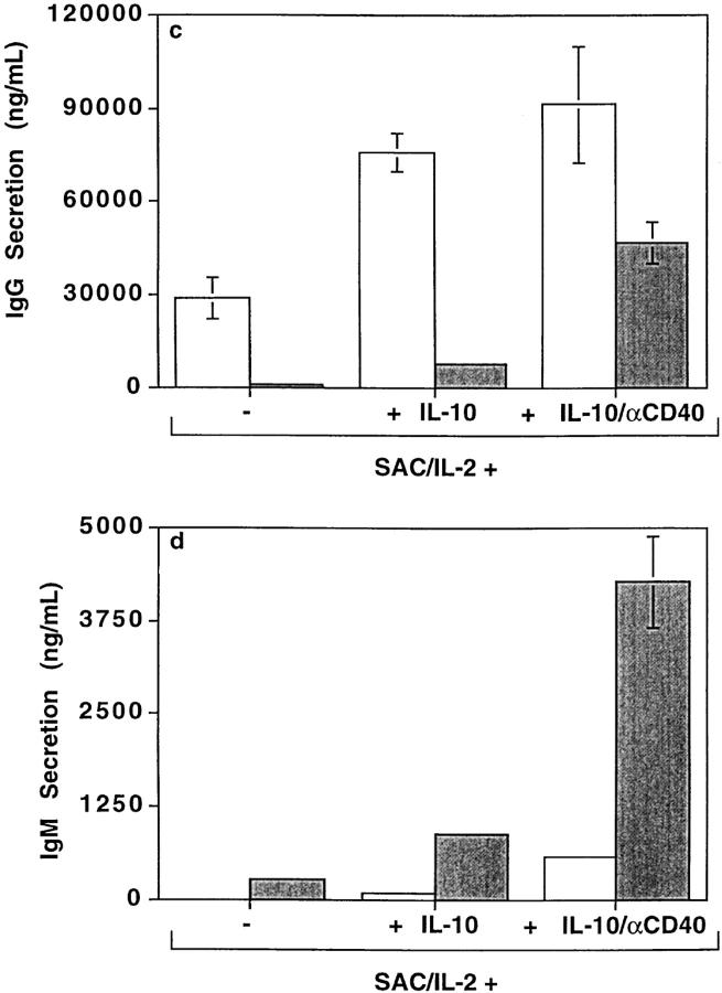
Memory B cells isolated from human tonsils are characterized by an activated cell surface phenotype, localization to mucosal epithelium, expression of somatically mutated immunoglobulin (Ig) variable (V) region genes, and a preferential differentiation into plasma cells in vitro. In spleens of both humans and rodents, a subset of memory B cells is believed to reside in the marginal zone of the white pulp. Similar to tonsil-derived memory B cells, splenic marginal zone B cells can be distinguished from naive follicular B cells by a distinct cell surface phenotype and by the presence of somatic mutations in their Ig V region genes. Although differences exist between human naive and memory B cells, no cell surface molecules have been identified that positively identify all memory B cells. In this study, we have examined the expression of the receptor-type protein tyrosine phosphatase CD148 on human B cells. CD148(+) B cells present in human spleen exhibited characteristics typical of memory B cells. These included an activated phenotype, localization to the marginal zone, the expression of somatically mutated Ig V region genes, and the preferential differentiation into plasma cells. In contrast, CD148(-) B cells appeared to be naive B cells due to localization to the mantle zone, the expression of surface antigens typical of unstimulated B cells, and the expression of unmutated Ig V region genes. Interestingly, CD148(+) B cells also coexpressed CD27, whereas CD148(-) B cells were CD27(-). These results identify CD148 and CD27 as markers which positively identify memory B cells present in human spleen. Thus, assessing expression of these molecules may be a convenient way to monitor the development of memory B cell responses in immunocompromised individuals or in vaccine trials.


Figures

Figure 1
Figure 1
CD148+ and CD148 human B cells represent phenotypically distinct subpopulations of B cells. (A) MNC from human spleen were incubated with PE-labeled anti-CD20 mAb and either FITC-labeled anti-CD148 mAb (bold histogram) or FITC-labeled isotype control IgG1 mAb (thin histogram). The FITC fluorescence of CD20+ B cells was assessed by flow cytometric analysis. The values represent the mean ± SD of CD148+ B cells from nine different donor spleens. (B) MNC from human spleen were incubated with FITC- or PE-labeled anti-CD148, PerCp-labeled anti-CD20, and one of the following PE- or FITC-labeled mAbs: anti-IgM, anti-IgD, anti-IgA, anti-CD21, anti-CD23, anti-CD27, anti-CD39, anti-CD80, anti-CD86, or anti-CD95. CD148+CD20+ (bold histogram) and CD148CD20+ (thin histogram) cells were gated, and the fluorescence and the light scattering characteristics of each population were assessed by flow cytometric analysis. The fluorescence of CD148+CD20+ and CD148CD20+ cells incubated with an isotype control mAb was within the first log of the histogram plot. These results are representative of six different spleen samples.
Figure 1
Figure 1
CD148+ and CD148 human B cells represent phenotypically distinct subpopulations of B cells. (A) MNC from human spleen were incubated with PE-labeled anti-CD20 mAb and either FITC-labeled anti-CD148 mAb (bold histogram) or FITC-labeled isotype control IgG1 mAb (thin histogram). The FITC fluorescence of CD20+ B cells was assessed by flow cytometric analysis. The values represent the mean ± SD of CD148+ B cells from nine different donor spleens. (B) MNC from human spleen were incubated with FITC- or PE-labeled anti-CD148, PerCp-labeled anti-CD20, and one of the following PE- or FITC-labeled mAbs: anti-IgM, anti-IgD, anti-IgA, anti-CD21, anti-CD23, anti-CD27, anti-CD39, anti-CD80, anti-CD86, or anti-CD95. CD148+CD20+ (bold histogram) and CD148CD20+ (thin histogram) cells were gated, and the fluorescence and the light scattering characteristics of each population were assessed by flow cytometric analysis. The fluorescence of CD148+CD20+ and CD148CD20+ cells incubated with an isotype control mAb was within the first log of the histogram plot. These results are representative of six different spleen samples.
Figure 2
Figure 2
Morphological differences between sort-purified human CD148+ and CD148 splenic B cells. Human splenic B cells were incubated with FITC-labeled anti-CD148 and PE-labeled anti-CD20 and sorted into CD20+ B cell populations that were either CD148 or CD148+. After sort-purification, the (A) CD148 B cells and (B) CD148+ B cells were cytocentrifuged onto glass slides and analyzed by Giemsa staining.
Figure 3
Figure 3
Localization of CD148+CD27+ and CD148CD27 B cells in follicles of human spleen. Serial tissue sections of human spleen were incubated with anti-IgD antiserum alone (blue; A and D), anti-IgD antiserum (blue) and anti-CD27 mAb (red; B and E), or anti-IgD antiserum (blue) and anti-IgM mAb (red; C). The anti-IgD polyclonal antibody was visualized after the addition of alkaline phosphatase–conjugated anti–goat Ig and phosphatase-specific substrate. The anti-CD27 or anti-IgM mAb was visualized by an anti–mouse Ig-specific Vectastain kit. Original magnification of A, B, and C: ×40; of D and E: ×100. The follicular mantle zone (FM), marginal zone (MZ), germinal center (G or GC), and T cell zones (T) are indicated.
Figure 4
Figure 4
Nucleotide sequence of Ig VH5 genes isolated from CD148 and CD148+ splenic B cells. Nucleotide sequences of VH5 Ig genes cloned from sort-purified CD148 and CD148+ human splenic B cells are shown. The germline VH5 (VH251) sequence was derived from Tomlinson et al. (reference 40). For clarity, only sequences homologous to VH251 are shown. Each dash represents identity with the germ-line sequence; nucleotide differences are indicated. The FR and CDRs are indicated.
Figure 4
Figure 4
Nucleotide sequence of Ig VH5 genes isolated from CD148 and CD148+ splenic B cells. Nucleotide sequences of VH5 Ig genes cloned from sort-purified CD148 and CD148+ human splenic B cells are shown. The germline VH5 (VH251) sequence was derived from Tomlinson et al. (reference 40). For clarity, only sequences homologous to VH251 are shown. Each dash represents identity with the germ-line sequence; nucleotide differences are indicated. The FR and CDRs are indicated.
Figure 5
Figure 5
Nucleotide sequence of Ig VH6 genes isolated from CD148 and CD148+ splenic B cells. Nucleotide sequences of VH6 Ig V genes cloned from sort-purified CD148 and CD148+ human splenic B cells are shown. The germline VH6 (VHVI) sequence was derived from Tomlinson et al. (reference 40). Each dash represents identity with the germ-line sequence; nucleotide differences are indicated. The FR and CDRs are indicated.
Figure 5
Figure 5
Nucleotide sequence of Ig VH6 genes isolated from CD148 and CD148+ splenic B cells. Nucleotide sequences of VH6 Ig V genes cloned from sort-purified CD148 and CD148+ human splenic B cells are shown. The germline VH6 (VHVI) sequence was derived from Tomlinson et al. (reference 40). Each dash represents identity with the germ-line sequence; nucleotide differences are indicated. The FR and CDRs are indicated.
Figure 6
Figure 6
Frequency of mutations in Ig V genes isolated from CD148 and CD148+ splenic B cells. The frequency of somatic mutations in (a) VH5 or (b) VH6 genes isolated from CD148 (white bars) and CD148+ B cells (black bars) was calculated as the number of nucleotide changes detected per base pairs sequenced. Each value represents the mean rate of mutation for either the entire VH5 or VH6 gene (VH), or for the different regions (FR, CDR) of the Ig V gene for all of the VH5 and VH6 clones analyzed.
Figure 7
Figure 7
CD148+ B cells differentiate into plasma cells in vitro. Sort-purified (a) CD148+ and (b) CD148 B cells were cultured for 3 d with anti-CD40 mAb (15 μg/ml), IL-2 (100 U/ml), and IL-10 (100 U/ml), and then recultured for an additional 3 d with IL-2 and IL-10. Plasma cell differentiation was then assessed by flow cytometric analysis after immunofluorescent staining with FITC–anti-CD38 mAb (x axis) and PE–anti-CD20 mAb (y axis). The values represent the percentage of cells in each quadrant of the dot plot. Similar results were obtained in a second experiment.
Figure 8
Figure 8
CD148 and CD148+ B cells differ in their ability to secrete Igs. Sort-purified CD148+ (gray bars) and CD148 B cells (white bars) were cultured with (a) anti-CD40 mAb (15 μg/ml) and IL-4 (400 U/ml), (b) anti-CD40 mAb and IL-2 (100 U/ ml) and IL-10 (100 U/ml), or (c and d) SAC (0.01%) plus IL-2 with or without IL-10 or anti-CD40 mAb. The amounts of IgG, IgE, IgG4, IgA, and IgM secreted were determined by isotype-specific immunoassays after 12 d. Each value represents the mean ± SEM of four to six replicates. The results shown are representative of data obtained from between 5 and 11 independent experiments.
Figure 8
Figure 8
CD148 and CD148+ B cells differ in their ability to secrete Igs. Sort-purified CD148+ (gray bars) and CD148 B cells (white bars) were cultured with (a) anti-CD40 mAb (15 μg/ml) and IL-4 (400 U/ml), (b) anti-CD40 mAb and IL-2 (100 U/ ml) and IL-10 (100 U/ml), or (c and d) SAC (0.01%) plus IL-2 with or without IL-10 or anti-CD40 mAb. The amounts of IgG, IgE, IgG4, IgA, and IgM secreted were determined by isotype-specific immunoassays after 12 d. Each value represents the mean ± SEM of four to six replicates. The results shown are representative of data obtained from between 5 and 11 independent experiments.

References

    1. MacLennan IC, Liu Y-J, Johnson GD. Maturation and dispersal of B-cell clones during T cell-dependent antibody responses. Immunol Rev. 1992;126:143–161. - PubMed
    1. Banchereau J, Rousset F. Human B lymphocytes: phenotype, proliferation, and differentiation. Adv Immunol. 1992;52:125–262. - PubMed
    1. Gray D. Immunological memory. Annu Rev Immunol. 1993;11:49–77. - PubMed
    1. Rajewsky K. Clonal selection and learning in the antibody system. Nature. 1996;381:751–758. - PubMed
    1. Liu Y-J, Grouard G, de Bouteiller O, Banchereau J. Follicular dendritic cells and germinal centers. Int Rev Cytol. 1996;166:139–179. - PubMed

Publication types

MeSH terms

Substances