. 1998 Dec 1;513 ( Pt 2)(Pt 2):611-9.
doi: 10.1111/j.1469-7793.1998.611bb.x.

Galvanic vestibular stimulation modulates voluntary movement of the human upper body

Affiliations

Galvanic vestibular stimulation modulates voluntary movement of the human upper body

A S Cauquil et al. J Physiol. .

Abstract

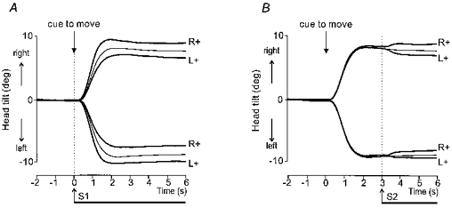
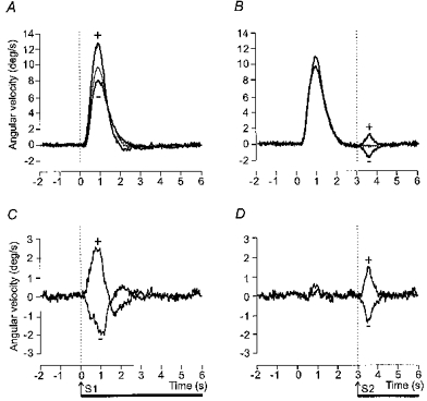
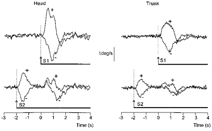
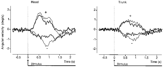
1. We have investigated whether vestibular information plays a role in the control of voluntary movement of the upper body. Movement consisted of a lateral tilt of the upper body in the frontal plane through an angle of about 8 deg. The influence of vestibular input was assessed from the effect of long duration (3-6 s), low-intensity (0.7 mA) galvanic vestibular stimulation (GVS) applied at different times relative to the movement. 2. GVS always produced a tilt of the body in the frontal plane but the response was larger and more prolonged when the onset of stimulation coincided with the cue to start moving compared with when it was applied some seconds after movement onset (i.e. while the subject was stationary in a tilted posture). 3. When the stimulus began 2 s before the voluntary movement the response consisted of two distinct components separated in time, one that was linked to the onset of GVS and another that was linked to onset of the voluntary movement. The large response observed when GVS onset coincided with the movement cue resembled the sum (after realignment in time) of these two separate components. 4. We suggest that these two components of the response to GVS relate to two different uses of vestibular information for whole-body control: first, to help maintain balance of the body, and second, to help guide and improve the accuracy of voluntary movements involving motion of the head in space.


Figures

Figure 1
Figure 1. Group mean head lateral tilt in space during voluntary movement and GVS
Superimposed movements to the right and left with the two polarities of stimulation, anode right (R+) or anode left (L+). Control movements without stimulation are shown by the thin traces. In A the onset of GVS coincided with the auditory cue to move. In B GVS was applied 3 s after the auditory cue when the movement was completed. Note that in both cases a given polarity of stimulation produced greater final tilt of the head in space for movements in one direction and smaller final tilt for movements in the opposite direction.
Figure 2
Figure 2. Group mean (±s.e.m.) angular displacement and velocity evoked by GVS
Shown are absolute values that have been calculated first by subtracting traces obtained in the control condition (no stimulation) from those obtained during stimulation. Angular displacement was measured 2 s after stimulus onset and peak angular velocity was measured over the same interval from the resulting difference traces. The contribution of each subject was taken as the mean absolute value obtained from each of the four conditions involving the two movement directions and two polarities of stimulation. Results are shown separately for the head and trunk, when GVS was applied either at the same time as the auditory cue to move (□, GVS with movement) or 3 s later (▪, GVS after movement).
Figure 3
Figure 3. Group mean head lateral angular velocity during voluntary movement and GVS
Upper panels (A and B) show superimposed the mean lateral angular velocity of the head in space for control movements (thin lines) and movements during which GVS was applied (thick lines). The movement cue was given at 0 s. Movements to the right and left have been averaged after inversion of traces from leftward movements. The trials in which GVS was applied have been sorted according to whether a given polarity of stimulation produced either greater deviation of the body in the direction of the movement (+) or less deviation (=) (see Fig. 1). Lower panels (C and D) show the same data after subtraction of the control movement (without stimulation) trace from the trials in which GVS was applied. In A and C GVS began at the same time as the movement cue. In B and D GVS began 3 s after the auditory cue when the movement was completed
Figure 4
Figure 4. Net effect of GVS on group mean head and trunk lateral angular velocity
Superimposed traces showing the net effect of the two polarities (+ and −; see Fig. 3 legend) of GVS, after subtraction of the control movement as in Fig. 3C and D, on the lateral angular velocity of the head (left panel) and trunk (right panel) in space. The traces have been realigned to the onset of stimulation at 0 s. GVS began either at the same time as the movement cue (thin lines) or 3 s after the cue when the movement was completed (thick lines).
Figure 5
Figure 5. Effect of starting GVS before the movement on the net GVS-evoked response
Superimposed traces showing the group mean net effect of the two polarities (+ and −; see Fig. 3 legend) of GVS, after subtraction of the control movement as in Fig. 3C and D, on the lateral angular velocity of the head (left traces) and trunk (right traces) in space. The movement cue was given at 0 s. GVS began either at the same time as the movement cue (upper traces) or 2 s before the cue when the body was stationary (lower traces). Note the two distinct components of the response when GVS began 2 s before the movement cue.
Figure 6
Figure 6. Artificial summation of the two components of the response obtained when GVS started before movement onset
Superimposed traces derived from data illustrated in Fig. 5 for the net GVS-evoked (after subtraction of control movement) lateral angular velocity of the head (left panel) and trunk (right panel) in space. The thin traces show the response when GVS began at the same time as the movement cue at 0 s. The thick traces have been artificially constructed from the two components of the response obtained when GVS began 2 s before the movement cue (see Fig. 5, lower traces). The first component of the response was shifted forward in time by 2 s and then summed with the second component of the response.

References

    1. Britton TC, Day BL, Brown P, Rothwell JC, Thompson PD, Marsden CD. Postural electromyographic response in the arm and the leg following galvanic vestibular stimulation in man. Experimental Brain Research. 1993;94:143–151. - PubMed
    1. Cauquil AS, Day BL. Influence of voluntary movement on the lateral tilt of body segments induced by galvanic vestibular stimulation in man. The Journal of Physiology. 1996;494.P:66P.
    1. Courjon JH, Precht W, Sirkin DW. Vestibular nerve and nuclei unit responses and eye movement responses to repetitive galvanic stimulation of the labyrinth in the rat. Experimental Brain Research. 1987;66:41–48. - PubMed
    1. Day BL, Cauquil AS, Bartolomei L, Pastor MA, Lyon IN. Human body-segment tilts induced by galvanic stimulation: a vestibularly driven balance protection mechanism. The Journal of Physiology. 1997;500:661–672. - PMC - PubMed
    1. Fitzpatrick R, Burke D, Gandevia SC. Task-dependent reflex responses and movement illusions evoked by galvanic vestibular stimulation in standing humans. The Journal of Physiology. 1994;478:363–372. - PMC - PubMed

Publication types

LinkOut - more resources