. 1998 Oct;28(10):2991-3002.
doi: 10.1002/(SICI)1521-4141(199810)28:10<2991::AID-IMMU2991>3.0.CO;2-B.

Transient alteration of T cell fine specificity by a strong primary stimulus correlates with T cell receptor down-regulation

Affiliations

Transient alteration of T cell fine specificity by a strong primary stimulus correlates with T cell receptor down-regulation

S Martin et al. Eur J Immunol. 1998 Oct.

Abstract

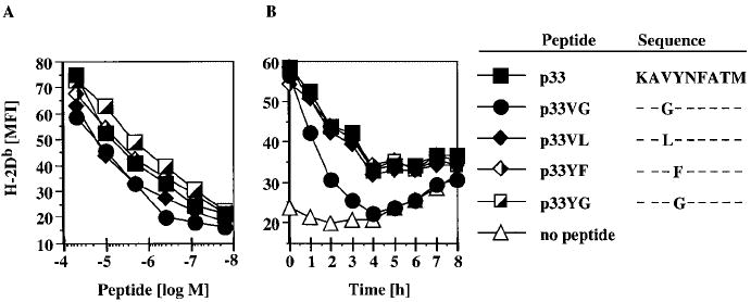
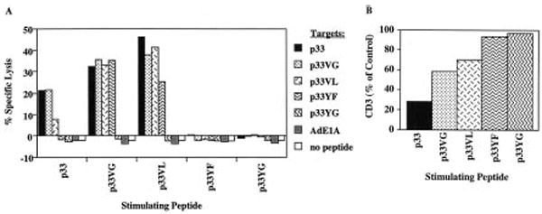
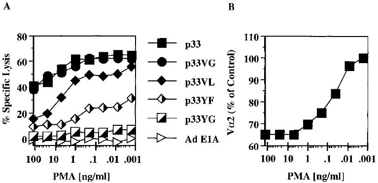
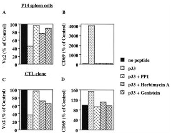
P14 mice expressing a transgenic TCR specific for the lymphocytic choriomeningitis virus glycoprotein p33 epitope were used to study the induction of CTL effector activity by a variety of ligands. Surprisingly, p33 variants which are weaker agonists for the P14 TCR than the wild-type p33 peptide were able to induce more potent effectors with a broader range of cytolytic specificity. Similarly, low concentrations of p33 were more effective than higher concentrations. These results correlated with no or only moderate TCR down-regulation by variants of p33 and low p33 concentrations. This phenotype observed after 18 h of culture was transient as progressive restoration of reactivity was observed at 42 or 66 h in the cultures stimulated with high p33 concentrations and this correlated with recovery of TCR surface levels. TCR down-regulation was blocked by src family kinase inhibitors. These findings indicate that the specificity of a T cell can be fine-tuned by the nature of the primary stimulus correlating with surface TCR level and imply an important role for src family kinases in the differential regulation of surface TCR levels upon TCR engagement by different ligand/MHC complexes.


Figures

Figure 1
Figure 1
H-2Db stabilization by p33 and its variants. (A) TAP-deficient RMA-S cells were preincubated overnight at 31 °C and graded concentrations of the indicated peptides added for 1 h before cells were shifted to 37 °C for 4 h. (B) Following overnight incubation at 31 °C, peptides were added to RMA-S cells at 20 μM for 1 h. BFA was then added for 2 h, peptide was washed away and the cells shifted to 37 °C. Cells were then incubated in the continuous presence of BFA and aliquots stained every hour. Staining was done with an H-2Db-specific antibody and cells analyzed by flow cytometry.
Figure 2
Figure 2
Agonist activity of p33 and its variants. (A) TCR transgenic P14 thymocytes were incubated with EL-4 cells as APC and graded concentrations of the indicated peptides for 18 h and then stained with anti-CD4 and anti-CD8. Deletion of immature CD4hiCD8hi DP thymocytes is given as % of control without peptide. (B) Graded concentrations of the indicated peptides were preincubated with 51Cr-labeled EL-4 cells for 30 min. P14.G5 CTL were then added and a standard 4-h Cr-release assay performed. The effector/target ratio was 5/1.
Figure 3
Figure 3
Different patterns of cytolysis are induced in P14 splenocytes by priming with wild-type or variant peptide. Correlation with surface CD3 level. TCR transgenic P14 spleen cells were stimulated for 18 h with EL-4 cells which had been prepulsed with 1 μM of the indicated peptides. (A) Cytotoxic activity was assayed after washing the cultures and addition of 51Cr-labeled, peptide-pulsed EL-4 target cells for 4 h at an E/T ratio of 100/1. (B) Corresponding CD3 levels on CD8+ cells of these cultures stimulated with the different peptides were determined by flow cytometry. Data are given as % MFI of control cells cultivated for 18 h with EL-4 cells without peptide.
Figure 4
Figure 4
Different patterns of cytolysis are induced in P14 splenocytes by stimulation with different concentrations of the agonist peptide p33 or p33VG. Correlation with surface CD3 level. P14 splenocytes were cultured for 18 h with irradiated EL-4 cells that had been pulsed with titered levels of p33, washed and labeled with fluorescent dye, BCECF-AM. Cultures were harvested, washed, and assayed immediately for lysis of 51Cr-labeled EL-4 pulsed with various peptides at 5 μM (A), and stained for surface CD3 levels on CD8+ cells (B). Alternatively, TCR transgenic P14 splenocytes were sorted away from BCECF-labeled EL-4 cells before assaying for cytolysis (C) and determination of surface CD3 level (D). E/T was 100/1. (E, F) P14 spleen cells were stimulated for 18 h with EL-4 cells that had been prepulsed with titered levels of p33VG. Lysis assays and determination of CD3 levels were done as for (A, B). No lysis was detected for 18-h cultures set up in the absence of peptide (not shown).
Figure 5
Figure 5
Desensitization of a CTL clone for cytotoxic reactivity by agonist pretreatment. Adherent MC57G cells were pulsed with the indicated concentrations of p33 for 1 h and washed three times. P14.G5 cells were added for 4 h, harvested and washed repeatedly. A 4-h Cr-release assay was performed using peptide-pulsed, 51Cr-labeled EL-4 target cells (A). Corresponding TCR Vα2 levels on the CTL were determined by flow cytometry at the time of harvest from the MC57G monolayers (B).
Figure 6
Figure 6
PMA-induced TCR down-regulation causes preferential loss of reactivity to weak agonists. P14.G5 cells were incubated for 1 h with the indicated concentrations of PMA. After three washes, peptide-pulsed EL-4 target cells were added and a 4-h 51Cr-release assay was performed (A). TCR Vα2 levels of CD8+ cells were determined by flow cytometry following the 1-h treatment with PMA (B).
Figure 7
Figure 7
Inhibition of protein tyrosine kinase activity blocks TCR down-regulation. P14 TCR transgenic spleen cells (A, B) or P14.G5 CTL clone (C, D) were preincubated for 60 min in the presence of PP1 or genistein. Preincubation with herbimycin A was done overnight with P14 spleen cells or for 60 min with P14.G5 CTL. EL-4 cells pulsed with p33 (1 μM) were then added for 4 h and cells stained for flow cytometry with anti-CD8 and anti-Vα2 antibodies (A, C) or with anti-CD8 and anti-CD69 antibodies (B, D). Mean fluorescence intensities are expressed as % or MFI of control in the absence of peptide.

References

    1. Sette A, Alexander J, Ruppert J, Snoke K, Franco A, Ishioka G, Grey HM. Antigen analogs/MHC complexes as specific T cell receptor antagonists. Annu Rev Immunol. 1994;12:413–431. - PubMed
    1. Jameson SC, Bevan MJ. T cell receptor antagonists and partial agonists. Immunity. 1995;2:1–11. - PubMed
    1. Sloan-Lancaster J, Allen PM. Altered peptide ligand-induced partial T cell activation: molecular mechanisms and role in T cell biology. Annu Rev Immunol. 1996;14:1–27. - PubMed
    1. Meier U-C, Klenermann P, Griffin P, James W, Köppe B, Larder B, McMichael A, Philipps R. Cytotoxic T lymphocyte lysis inhibited by viable HIV mutants. Science. 1995;270:1360–1362. - PubMed
    1. Bertoletti A, Sette A, Chisari FV, Penna A, Levrero M, De Carli M, Fiaccadori F, Ferrari C. Natural variants of cytotoxic epitopes are T-cell receptor antagonists for antiviral cytotoxic T cells. Nature. 1994;369:407–410. - PubMed

Publication types