. 1998 Nov 1;12(21):3369-81.
doi: 10.1101/gad.12.21.3369.

JNKK1 organizes a MAP kinase module through specific and sequential interactions with upstream and downstream components mediated by its amino-terminal extension

Affiliations

JNKK1 organizes a MAP kinase module through specific and sequential interactions with upstream and downstream components mediated by its amino-terminal extension

Y Xia et al. Genes Dev. .

Abstract

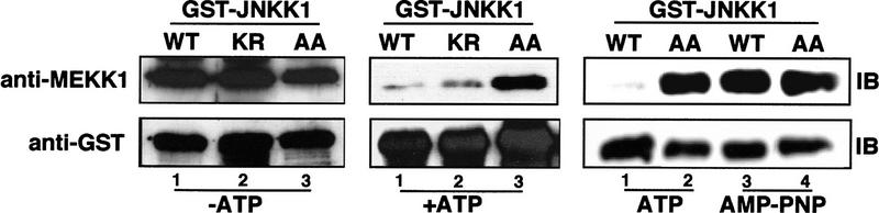
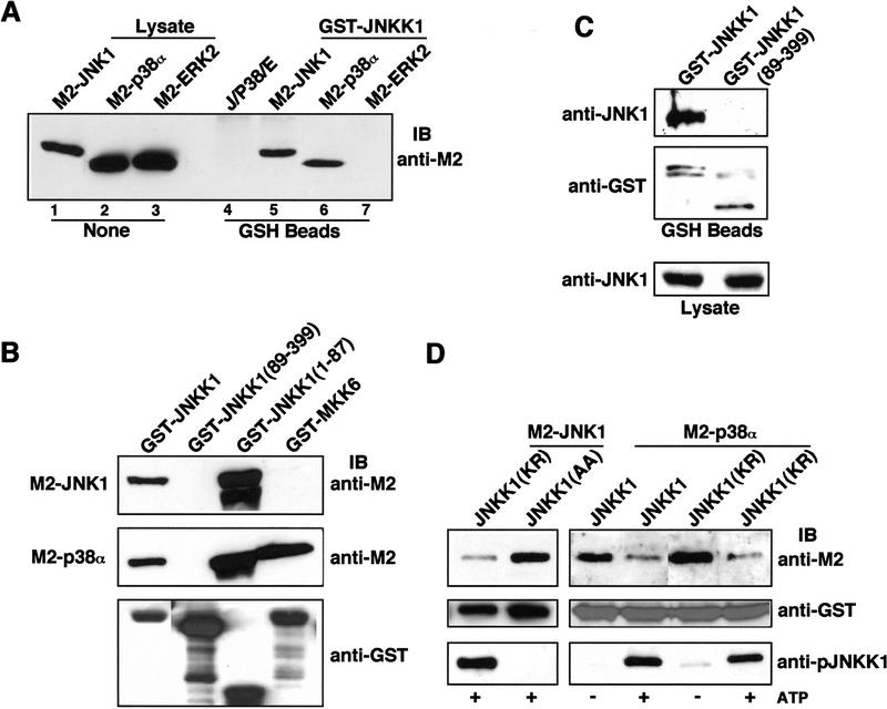
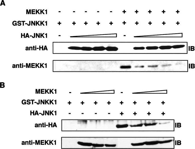
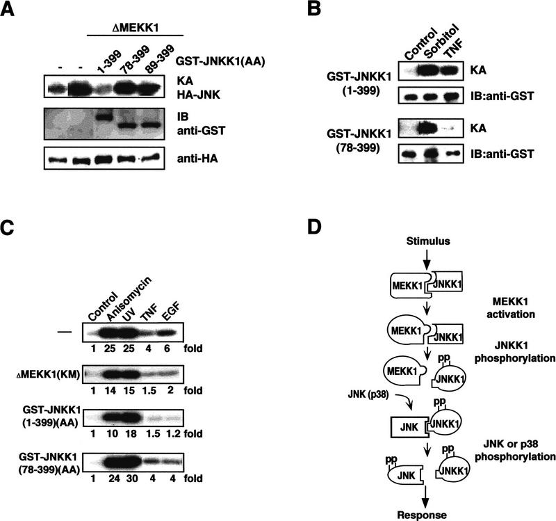
MAP kinase (MAPK) cascades are composed of a MAPK, MAPK kinase (MAPKK), and a MAPKK kinase (MAPKKK). Despite the existence of numerous components and ample opportunities for crosstalk, most MAPKs are specifically and distinctly activated. We investigated the basis for specific activation of the JNK subgroup of MAPKs. The specificity of JNK activation is determined by the MAPKK JNKK1, which interacts with the MAPKKK MEKK1 and JNK through its amino-terminal extension. Inactive JNKK1 mutants can disrupt JNK activation by MEKK1 or tumor necrosis factor (TNF) in intact cells only if they contain an intact amino-terminal extension. Mutations in this region interfere with the ability of JNKK1 to respond to TNF but do not affect its activation by physical stressors. As JNK and MEKK1 compete for binding to JNKK1 and activation of JNKK1 prevents its binding to MEKK1, activation of this module is likely to occur through sequential MEKK1:JNKK1 and JNKK1:JNK interactions. These results underscore a role for the amino-terminal extension of MAPKKs in determination of response specificity.


Figures

Figure 1
Figure 1
MEKK1 selectively activates JNK and p38. Cos-1 cells were transiently transfected with expression vectors for HA-tagged JNK1 (A,D), p38α (B,E), or ERK2 (C,F), together with increasing amounts (in ng) of either ΔMEKK1 (A,B,C) or FL-MEKK1 (D,E,F) vectors. After 48 hr, 10-μg samples of transfected cell lysates were used to determine MAPK expression by immunoblotting with anti-HA (bottom panels). Fifty-microgram samples were subjected to immunecomplex kinase assays using anti-HA and appropriate recombinant proteins as substrates (top panels). Fold stimulations of MAPK activities were calculated after phosphoimaging and normalization for MAPK expression levels. As a reference, JNK1 was stimulated by UV irradiation (40 J · m−2), p38α by UV irradiation or anisomycin (1 μg/ml), and ERK2 by treatment with phorbol ester (TPA, 10 ng/ml).
Figure 2
Figure 2
JNKK1 is the preferred MAPKK substrate for MEKK1. (A) MEKK1 preferentially activates JNKK1 in mammalian cells. Cos-1 cells were transiently transfected with increasing amounts of ΔMEKK1, together with HA-tagged MAPKKs. Immunoblot analyses and immunecomplex kinase assays were performed as described in Fig. 1, using recombinant, catalytically inactive MAPKs as substrates. Fold stimulations of MAPKK activities were calculated as in Fig. 1 and are indicated below each autoradiograph. As references, JNKK1, MKK3, and MKK6 activities were stimulated by UV irradiation of transfected cells, and MEK1/2 by TPA treatment. (B) JNKK1 is the preferred MAPKK substrate for MEKK1 in vitro. Kinase assays (see Materials and Methods) were performed using purified recombinant GST–ΔMEKK1 as the kinase and purified recombinant GST–MAPKK fusion proteins, as well as JNKK1 truncation mutants—JNKK1(78–399) or JNKK1(89–399)—as substrates. Incorporation of 32P was determined by scintillation counting. Data were corrected for MAPKK autophosphorylation and fit to the Michaelis–Menten equation. (Inset) The substrate specificity constants (Vmax/Km).
Figure 3
Figure 3
JNKK1 specifically interacts with MEKK1. (A) JNKK1, but not MKK6, forms stable complexes with MEKK1. Lysates of Cos-1 cells cotransfected with the indicated amounts of ΔMEKK1 and either HA–JNKK1 or HA–MKK6b expression vectors were immunoprecipitated (IP) with anti-HA. The precipitates were separated by SDS-PAGE and analyzed by immunoblotting (IB) with anti-MEKK1. The efficiencies of HA–JNKK1 and HA–MKK6 expression were similar (data not shown). (B) JNKK1, but not other MAPKKs, interacts with MEKK1. Lysates of Cos-1 cells transiently expressing HA–MAPKKs were mixed with lysates containing ΔMEKK1. The mixtures were immunoprecipitated with anti-HA, the immunocomplexes were separated by SDS-PAGE and immunoblotted sequentially with anti-MEKK1 (top) and anti-HA (bottom). (C) JNKK1, but not JNKK2, interacts with MEKK1. Cos-1 cells were transiently transfected with ΔMEKK1 and either HA–JNKK1, HA–JNKK2, or empty expression vector. Cell lysates were either directly separated by SDS-PAGE or immunoprecipitated with anti-HA as indicated and then resolved by SDS-PAGE. Immunoblot analyses were performed with anti-MEKK1 (top) or anti-HA (bottom). (D) Raf-1 interacts with MEK2 but not with JNKK1. Cos-1 cells were transfected with Raf1(BXB), HA–JNKK1, and HA–MEK2 expression vectors, as indicated. Cell lysates were analyzed as described in A. Protein expression was evaluated by separating one-fortieth of each lysate directly by SDS-PAGE and immunoblotting with anti-Raf-1 (top) or anti-HA (bottom) antibodies. The same antibodies were used for immunoblotting of anti-HA immunoprecipitates. (E) Amino-terminal extension of JNKK1 is required for interacting with MEKK1 and JNKs(p38). Schematic representation of JNKK1 deletion mutants with either GST or HA tags at their amino termini. The location of the ATP-binding site is indicated by an asterisk and the activating phosphoacceptor sites by pp. GST-tagged full-length and truncated JNKK1 proteins were expressed either in Cos-1 cells or in bacteria and their binding to ΔMEKK1, JNK, and p38 was examined by in-vitro mixing–coprecipitation assays, as described in B. The results are indicated on the right.
Figure 4
Figure 4
MEKK1 binds to JNKK1. (A) Full-length MEKK1 specifically interacts with JNKK1 through its carboxy-terminal kinase domain. GST–MAPKK fusion proteins were expressed in E. coli and purified. Equal amounts of each protein were mixed with lysates of transfected Cos-1 cells containing transiently expressed wild-type (WT) or catalytically inactive (KM) MEKK1 with an amino-terminal Express epitope. The proteins were precipitated with GSH–Sepharose and analyzed by immunoblotting with anti-Express (top), anti-MEKK1 directed against the carboxy-terminal kinase domain (middle), or anti-GST (bottom). (B) Purified recombinant JNKK1 specifically and directly interacts with MEKK1. Purified, recombinant GST–MAPKK fusion proteins were mixed with lysates of cells expressing ΔMEKK1 (top) or with purified His–ΔMEKK1 expressed in E. coli (middle). The proteins were precipitated with GSH–Sepharose and analyzed by immunoblotting with anti-MEKK1 or anti-GST (bottom). (C) Binding of JNKK1 to purified ΔMEKK1. Purified recombinant GST–ΔMEKK1 was mixed with Cos-1 cell lysates containing equal amounts of transiently expressed HA–JNKK1, HA–MEK1, or HA–MEK2 determined by anti-HA immunoblotting (bottom). The proteins were precipitated with GSH–Sepharose and analyzed by immunoblotting with anti-HA (top) or anti-GST (middle).
Figure 5
Figure 5
Phosphorylation of JNKK1 disrupts its binding to ΔMEKK1. GST-fusion proteins containing wild-type JNKK1, an ATP-binding mutant, JNKK1 (KR), or a mutant lacking the activating phosphoacceptor sites, JNKK1(AA), were transiently expressed in Cos-1 cells. Lysates containing these proteins were mixed with ΔMEKK1-containing lysates and incubated for 20 min at 30°C in kinase buffer without ATP or with 1 mm ATP or AMP–PNP as indicated. Next, the GST fusion proteins were precipitated with GSH–Sepharose. After washing six to eight times, the precipitates were resolved by SDS-PAGE and immunoblotted with anti-MEKK1 (top) and anti-GST (bottom).
Figure 6
Figure 6
JNKK1 specifically interacts with JNK1 and p38α. (A) JNKK1 stably interacts with JNK1 and p38α, but not with ERK2. Lysates (10 μg) of Cos-1 cells transfected with M2-tagged MAPK vectors were examined for MAPK expression by immunoblotting with anti-M2 (lanes 1–3). Lysates (400 μg) were also mixed with lysates containing GST–JNKK1, precipitated with GST–Sepharose, and immunoblotted with anti-M2. In addition, equal amounts of lysates containing each of the MAPKs were mixed and examined for binding to GSH–Sepharose in the absence of GST–JNKK1 (lane 4). (B) JNKK1 interacts with both JNK1 and p38α through its amino-terminal extension. Lysates containing M2–JNK1 or M2–p38α were mixed with purified, recombinant GST–JNKK1, GST–JNKK1(89–399), GST–JNKK1(1–87), or GST–MKK6. After precipitation with GSH–Sepharose and separation by SDS-PAGE, the precipitated proteins were immunoblotted with anti-M2 (top two panels) or anti-GST (bottom panel). (C) JNKK1 binds endogenous JNK1. HEK293 cells were transiently transfected with either GST–JNKK1 or GST–JNKK1(89–399) expression vectors. Cell lysates were prepared, precipitated with GSH–Sepharose, separated by SDS-PAGE, and immunoblotted with anti-JNK1 or anti-GST. A small fraction (formula image) of each lysate was directly analyzed for its content of JNK1. (D) Lysates of Cos-1 cells expressing GST–JNKK1, GST–JNKK1(KR), or GST–JNKK1(AA) were incubated with a small amount of lysate containing ΔMEKK1 in kinase buffer in the absence or presence of 1 mm ATP. Then, lysates containing similar amounts of M2–JNK1 (left panels) or M2–p38α (right panels) were added. The mixtures were precipitated with GSH–Sepharose followed by immunoblotting with anti-M2 (top panels), anti-GST (middle panels), or anti-phospho-JNKK1 (bottom panels).
Figure 7
Figure 7
ΔMEKK1 and JNK compete for binding to JNKK1. (A) Cell lysates containing GST–JNKK1 or a mixture of GST–JNKK1 lysate with ΔMEKK1-containing lysate were mixed with increasing amounts of HA–JNK1-containing lysates. The mixtures were precipitated with GSH–Sepharose, resolved by SDS-PAGE, and immunoblotted with anti-HA (top) and anti-MEKK1 (bottom). (B) Lysates containing GST–JNKK1 were mixed with increasing amounts of ΔMEKK1-containing lysates in the absence or presence of HA–JNK1-containing lysates. The mixtures were precipitated with GSH–Sepharose and analyzed as described above.
Figure 8
Figure 8
The amino-terminal extension of JNKK1 is required for transducing upstream stimuli to JNK in intact cells. (A) The amino-terminal extension is required for inhibition of JNK activation by dominant-negative JNKK1. HeLa cells were cotransfected with HA–JNK2 and ΔMEKK1 expression vectors, along with either empty expression vector (−) or expression vectors for FL(1–399) GST–JNKK1(AA) or amino-terminally truncated (78–399 and 89–399) GST–JNKK1(AA). After 48 hr, JNK activity was determined by immunecomplex kinase assays (KA) using anti-HA antibody and GST–c-Jun(1–79) as a substrate (top panel). The content of GST–JNKK1(AA) proteins and HA–JNK2 was determined by immunoblotting (IB; bottom two panels). (B) The amino-terminal extension of JNKK1 is required for response to stimuli. HeLa cells were transfected with expression vectors for either FL(1-399) or amino-terminally truncated (78–399) GST–JNKK1. After 48 hr, the cells were incubated with either 0.4 m sorbitol (osmotic shock) or 20 ng/ml of TNF for 20 min. Cell lysates were prepared, the GST–JNKK1 proteins were precipitated with GSH–Sepharose, and their kinase activities were determined. (C) The amino-terminal extension of JNKK1 is required for inhibition of JNK activation in response to physiological stimuli. HeLa cells were cotransfected with HA–JNK2 and either an empty vector (−) or expression vectors for catalytically inactive ΔMEKK1 [MEKK1(KM)] and either FL(1–399) or amino-terminally truncated (78–399) GST–JNKK1(AA). After 40 hr, the cells were exposed to anisomycin (15 ng/ml), UV-C (20 J · m−2), TNF (5 ng/ml), or EGF (20 ng/ml). Lysates were prepared after 20 min and HA–JNK2 kinase activity was determined as described above. (D) A sequential-interaction model for organization of the MEKK1–JNKK1–JNK(p38) MAPK module. MEKK1, either active or inactive, interacts with inactive JNKK1 to form a MEKK1:JNKK1 complex, whose formation depends on the amino-terminal extension of JNKK1. Activated MEKK1 phosphorylates and activates JNKK1, resulting in dissociation of the MEKK1:JNKK1 complex. Activated JNKK1 then interacts with JNK (or p38) through its amino-terminal extension. JNK (or p38) activation is followed by dissociation of the JNKK1:JNK complex and activated JNK is freed to bind its targets and phosphorylate them.

References

    1. Avruch J, Zhang X, Kyriakis J. Raf meets ras: Completing the framework of a signal transduction pathway. Trends Biochem Sci. 1994;19:279–283. - PubMed
    1. Bardwell L, Cook J, Chang E, Cairns B, Thorner J. Signaling in the yeast pheromone response pathway: Specific and high-affinity interaction of the mitogen-activated protein (MAP) kinases Kss1 and Fus3 with the upstream MAP kinase kinase Ste7. Mol Cell Biol. 1996;16:3637–3650. - PMC - PubMed
    1. Bax B, Jhoti H. Protein-protein interactions: putting the pieces together. Curr Biol. 1995;5:1119–1121. - PubMed
    1. Blank JL, Gerwins P, Elliott EM, Sather S, Johnson GL. Molecular cloning of mitogen-activated protein/ERK kinase kinases (MEKK) 2 and 3. J Biol Chem. 1996;274:5361–5368. - PubMed
    1. Brewster JL, de Valoir T, Dwyer ND, Winter E, Gustin MC. An osmosensing signal transduction pathway in yeast. Science. 1993;259:1760–1763. - PubMed

Publication types

MeSH terms

Substances

Associated data