. 1998 Nov 1;12(21):3431-41.
doi: 10.1101/gad.12.21.3431.

Bacterial senescence: stasis results in increased and differential oxidation of cytoplasmic proteins leading to developmental induction of the heat shock regulon

Affiliations

Bacterial senescence: stasis results in increased and differential oxidation of cytoplasmic proteins leading to developmental induction of the heat shock regulon

S Dukan et al. Genes Dev. .

Abstract

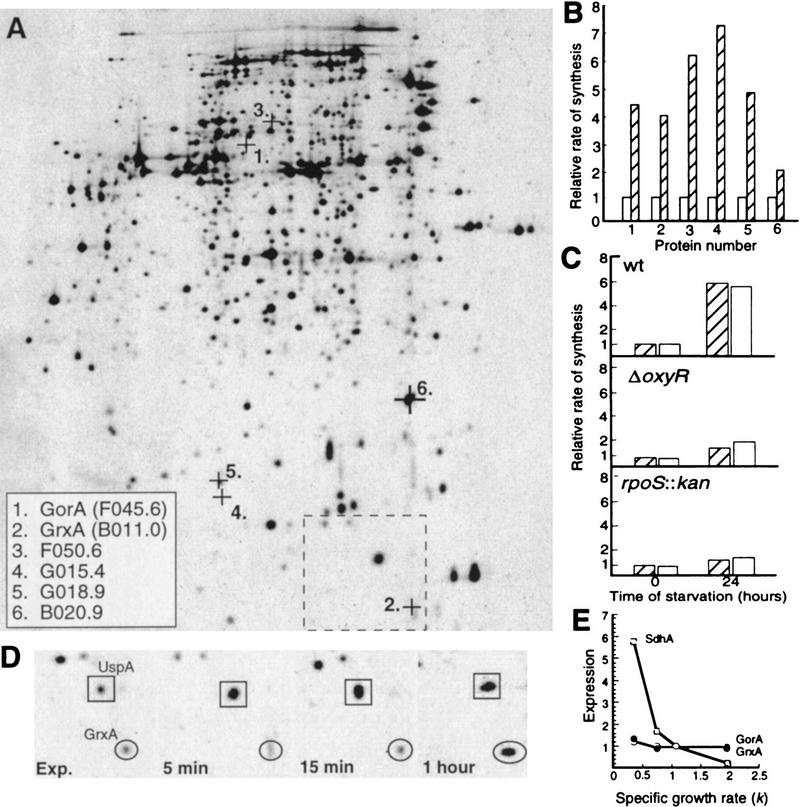
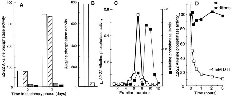
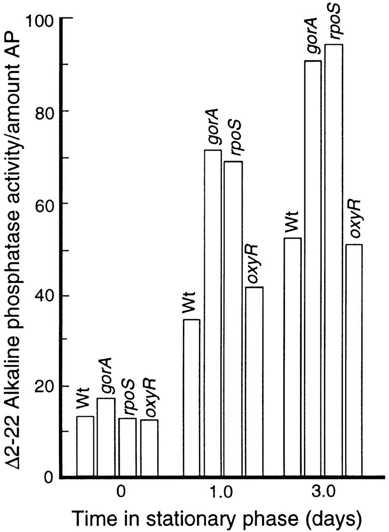
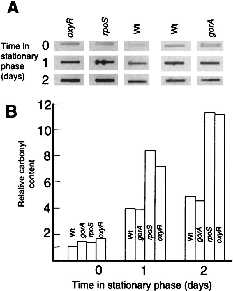
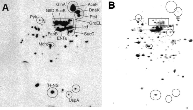
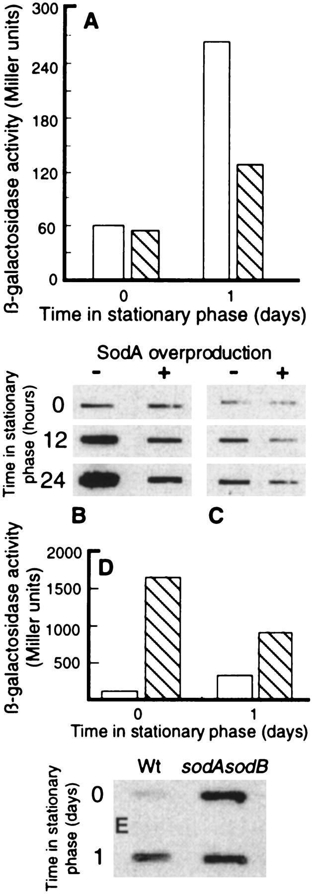
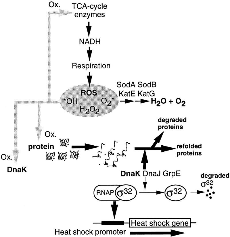
Aging, or senescence, is the progressive deterioration of every bodily function over time. A fundamental question that applies to all life forms, including growth-arrested bacteria, is why growing older by necessity causes organisms to grow more fragile. In this work, we demonstrate that the levels of oxidized proteins is correlated to the age of a stationary-phase Escherichia coli culture; both disulfide bridge formation of a cytoplasmic leader-less alkaline phosphatase and protein carbonyl levels increase during stasis. The stasis-induced increase in protein oxidation is enhanced in cells lacking the global regulators OxyR and sigmas. Some proteins were found to be specifically susceptible to stasis-induced oxidation; notably several TCA cycle enzymes, glutamine synthetase, glutamate synthase, pyruvate kinase, DnaK, and H-NS. Evidence that oxidation of target proteins during stasis serves as the signal for stationary-phase, developmental, induction of the heat shock regulon is presented by demonstrating that this induction is mitigated by overproducing the superoxide dismutase SodA. In addition, cells lacking cytoplasmic superoxide dismutase activity exhibit superinduction of heat shock proteins. The possibility that oxidative sensitivity of TCA cycle enzymes serves as a feedback mechanism down-regulating toxic respiration is discussed.


Figures

Figure 1
Figure 1
Synthesis of oxidative-defense proteins during starvation. (A) Location on the two-dimensional reference gel of the OxyR-dependent oxidative-stress proteins identified as inducible by carbon starvation. The alpha-numeric designations and protein names of identified proteins are shown in the inset box. The box with broken lines denotes the area shown in D. (B) Relative rate of synthesis of oxidative-stress proteins during growth (OD = 0.4 ± 0.05) (open bars) and after 16 hr carbon starvation (hatched bars). The rate of synthesis during exponential growth was assigned a value of 1.0. (C) Relative rate of GorA (hatched bars) and GrxA (open bars) synthesis during growth and starvation in wild-type, ΔoxyR, and rpoS::kan backgrounds. The rate of synthesis of GorA and GrxA during exponential growth in the wild-type background was assigned a value of 1.0. (D) Kinetics of GrxA synthesis in comparison to UspA during carbon starvation. (E) Relative rates of synthesis of GorA (•) and GrxA (○) in comparison to SdhA (□) at varying growth rates. The cultures were grown exponentially in minimal MOPS media supplemented with acetate, glycerol, or glucose as carbon sources and glucose plus amino acids, nucleotides, and vitamins for rich medium. The rates of protein synthesis are plotted relative to the rate of synthesis in glucose minimal media which was assigned a value of 1.0. Growth rates in the different media are expressed as k, the first-order growth-rate constant. The analysis was repeated three times to confirm reproducibility. Representative results are presented in the figure and the standard deviation was always <10% in the measurements of rates of individual protein synthesis.
Figure 2
Figure 2
(A) Δ2-22AP activity during growth (time zero) and in stationary phase (2 days). AP activity measured in aerobically grown cells was normalized to total cell mass (OD600) before (open bars) and after an osmotic shock treatment (hatched bars). Activity was measured also in cells grown and starved anaerobically (shaded bars) and aerobically in the presence of 4 mm DTT (black bars). (B) Wild-type AP activity before (open bars) and after an osmotic shock (hatched bars). (C) Determination of Δ2-22AP activity after separation of crude extract by gel filtration on Sephacryl HR 100. The relative levels of Δ2-22AP in the eluted fractions were determined by Western blotting using polyclonal antibodies against AP and quantification of Δ2-22AP bands was performed electronically using the ImageQuant (Molecular Dynamics) software. (D) Effect of DTT (4 mm) on the in vitro Δ2-22 AP activity. See text for details. The analysis was repeated three times to confirm reproducibility. Representative results are presented and the standard deviation was always <7% in the measurements of AP activity.
Figure 3
Figure 3
Δ2-22AP activity in wild-type, and gorA, oxyR, and rpoS mutants during growth (time zero) and stationary phase. All activity measurements were normalized to Δ2-22AP levels determined by Western blotting as described in Materials and Methods. The analysis was repeated three times to confirm reproducibility. The standard deviation was <8%.
Figure 4
Figure 4
(A) Autoradiograph showing protein carbonyl levels in wild-type cells and different mutants, as indicated, during growth (time zero, OD = 0.5 ± 0.05) and stationary phase (1 and 2 days). Equal amounts of protein were loaded in each slot. (B) Quantification of carbonyl levels using the ImageQuant software (Molecular Dynamics). The analysis was repeated three times to confirm reproducibility. Representative results are presented.
Figure 5
Figure 5
Specific protein carbonylation determined by two-dimensional Western blot immunoassay. (A) An autoradiograph obtained after carbonyl immunoassay of proteins from a wild-type E. coli culture starved for 1 day; (B) the same protein extract blotted to PVDF membrane and stained with Coomassie brilliant blue. (GltD) Glutamate synthase; (GlnA) glutamine synthetase; (Icd) isocitrate dehydrogenase; (SucB) dihydrolipoamide succinyltransferase; (Mdh) malate dehydrogenase; (AceF) dihydrolipoamide acetyltransferase; (SucC) succinyl CoA ligase; (PtsI) phosphoenolpyruvate–protein phosphotransferase; (Pyk) pyruvate kinase; (UspA) universal stress protein A; (FabB) β-ketoacyl-[acyl carrier protein] synthetase; (EF-Tu) elongation factor Tu. The analysis was repeated two times to confirm reproducibility. Representative results are presented.
Figure 6
Figure 6
Effects of overproducing SodA on stasis-induced heat shock gene expression. (A) groEL promoter activity in the absence (open bars) and presence (hatched bars) of elevated SodA levels during growth (time zero, OD = 0.3 ± 0.05) and in stationary phase (day 1). Expression of GroEL was determined by measuring β-galactosidase activity in cells harboring a chromosomal groEL–lacZ fusion. (B) Levels of DnaK during growth (time zero) and at times in stationary phase in the absence or presence of elevated levels of SodA. The levels of DnaK was determined by Western blot analysis using monoclonal mouse anti-DnaK antibodies. (C) Protein carbonyl levels during growth (time zero) and at times in stationary phase in the absence or presence of elevated levels of SodA. Equal amounts of protein were loaded in each slot. (D) groEL promoter activity in the wild-type strain (open bars) and a sodA sodB double mutant (hatched bars) during growth (time zero, OD = 0.3 ± 0.05) and stationary phase (1 day). (E) Levels of DnaK during growth (time zero) and after 1 day of stationary phase in wild-type and sodA sodB double mutant strains. The levels of DnaK was determined by Western blot analysis using monoclonal mouse anti-DnaK antibodies. The analysis was repeated three times (Miller units) or two times (Western blot) to confirm reproducibility. Representative results are presented in the figure and the standard deviation was always <10%.
Figure 7
Figure 7
Schematic representation of the model for stasis-induced, developmental, induction of the heat shock regulon. The model is very much the same as the proposed signal-response transduction pathway for heat shock induction presented by Bukau (1993) with the addition of DnaJ, DnaK, and GrpE, as negative modulators of the response (for review, see Bukau 1993; Gross 1996). Central to this model is that the heat shock proteins DnaJ, DnaK, and GrpE, have dual functions; one is to aid refolding of denatured or misfolded proteins and the other is to promote proteolytic degradation of the heat shock regulator σ32. An increase in the cellular level of denatured proteins as a consequence of heat or ethanol treatment will sequester the DnaJ, DnaK, and GrpE components (provided at least one of them is limiting) through binding to their denatured substrate. This will allow stabilization of σ32, which will trigger increased transcription of the heat shock genes (e.g., Bukau 1993). During stasis, we propose that oxidative damage of target proteins will similarly sequester these heat shock modulators allowing induction of the regulon. This notion is supported by the fact that heat shock induction is markedly suppressed by overproducing SodA during stasis. In addition, the fact that DnaK appears to be specifically sensitive to oxidative carbonylation opens up the possibility that oxidative damage of DnaK itself may be involved in stasis-induced heat shock gene expression. Presumably, oxidation of DnaK will destroy or reduce its ability to participate in the degradation of σ32 which, in turn, will activate heat shock promoters.

References

    1. Albertson NH, Nyström T. Effects of starvation for exogenous carbon on functional mRNA stability and rate of peptide chain elongation in Escherichia coli. FEMS Microbiol Lett. 1994;117:181–187. - PubMed
    1. Almiron M, Link AJ, Furlong D, Kolter R. A novel DNA-binding protein with regulatory and protective roles in starved Escherichia coli. Genes & Dev. 1992;6:2646–2654. - PubMed
    1. Amici A, Levine RL, Tsai L, Stadtman ER. Conversion of amino acid residues in proteins and amino acid homopolymers to carbonyl derivatives by metal-catalyzed oxidation reactions. J Biol Chem. 1989;264:3341–3346. - PubMed
    1. Becker-Hapak M, Eisenstark A. Role of rpoS in the regulation of glutathione oxidoreductase (gor) in Escherichia coli. FEMS Microbiol Lett. 1995;134:39–44. - PubMed
    1. Benov L, Fridovich I. A superoxide dismutase mimic protects sodA sodB Escherichia coli against aerobic heating and stationary-phase death. Arch Biochem Biophys. 1995;322:291–294. - PubMed

Publication types

MeSH terms

LinkOut - more resources