

Cell type-specific expression of angiopoietin-1 and angiopoietin-2 suggests a role in glioblastoma angiogenesis
- PMID: 9811337
- PMCID: PMC1853417
- DOI: 10.1016/S0002-9440(10)65733-1
Cell type-specific expression of angiopoietin-1 and angiopoietin-2 suggests a role in glioblastoma angiogenesis
Abstract
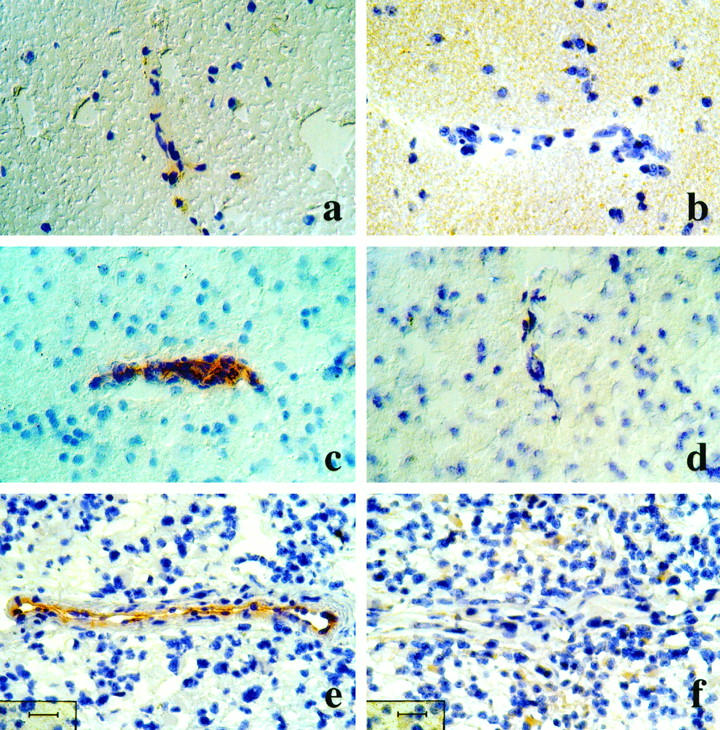
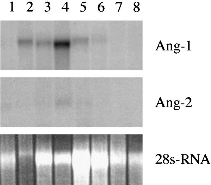
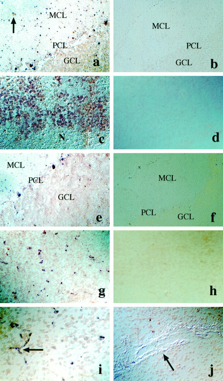
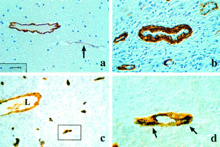
Glioblastomas are highly vascular tumors which overexpress the angiogenesis factor vascular endothelial growth factor (VEGF). VEGF and its receptors, VEGF-R1 and VEGF-R2, have been shown to be necessary for embryonic angiogenesis as well as for tumor angiogenesis. Recently, the angiopoietin/Tie2 receptor system has been shown to exert functions in the cardiovascular system that are distinct from VEGF but are also critical for normal vascular development. To assess the potential role of Tie2 and its ligands angiopoietin-1 and angiopoietin-2 in tumor vascularization, we analyzed their expression pattern in human gliomas. Tie-2 was up-regulated in tumor endothelium compared to normal human brain tissue. We further observed cell type-specific up-regulation of the message for both angiopoietin-1 and angiopoietin-2 in gliomas. Whereas Ang-1 mRNA was expressed in tumor cells, Ang-2 mRNA was detected in endothelial cells of a subset of glioblastoma blood vessels. Small capillaries with few periendothelial support cells showed strong expression of Angiopoietin-2, whereas larger glioblastoma vessels with many periendothelial support cells showed little or no expression. Although the function of Tie2 and its ligands in tumor angiogenesis remains a subject of speculation, our findings are in agreement with a recently proposed hypothesis that in the presence of VEGF, local production of Ang-2 might promote angiogenesis.
Figures
Comment in
-
Is angiopoietin-2 necessary for the initiation of tumor angiogenesis?Am J Pathol. 1998 Nov;153(5):1333-9. doi: 10.1016/S0002-9440(10)65717-3. Am J Pathol. 1998. PMID: 9811321 Free PMC article. Review. No abstract available.
References
-
- Kleihues P, Burger PC, Plate KH, Ohgaki H, Cavenee WK: Glioblastoma: Pathology and Genetics of Tumours of the Nervous System. Edited by Kleihues P, Cavenee WK. Lyon, France, International Agency for Research on Cancer, 1997, pp 16–24
-
- Brem S, Cotran R, Folkman J: Tumor angiogenesis: a quantitative method for histological grading. J Natl Cancer Inst 1972, 48:347-356 - PubMed
-
- Folkman J: Tumor angiogenesis. Adv Cancer Res 1985, 43:175-203 - PubMed
-
- Risau W: Mechanisms of angiogenesis. Nature 1997, 386:671-674 - PubMed
-
- Carmeliet P, Ferreira V, Breier G, Pollefeyt S, Kieckens L, Gertsenstein M, Fahrig M, Vandenhoeck A, Harpal K, Eberhardt C, Declercq C, Pawling J, Moons L, Collen D, Risau W, Nagy A: Abnormal blood vessel development and lethality in embryos lacking a single VEGF allele. Nature 1996, 383:73-75 - PubMed
Publication types
MeSH terms
Substances
LinkOut - more resources
Full Text Sources
Other Literature Sources
Miscellaneous
