

Chromosomal imbalances in papillary renal cell carcinoma: genetic differences between histological subtypes
- PMID: 9811338
- PMCID: PMC1853413
- DOI: 10.1016/S0002-9440(10)65734-3
Chromosomal imbalances in papillary renal cell carcinoma: genetic differences between histological subtypes
Abstract
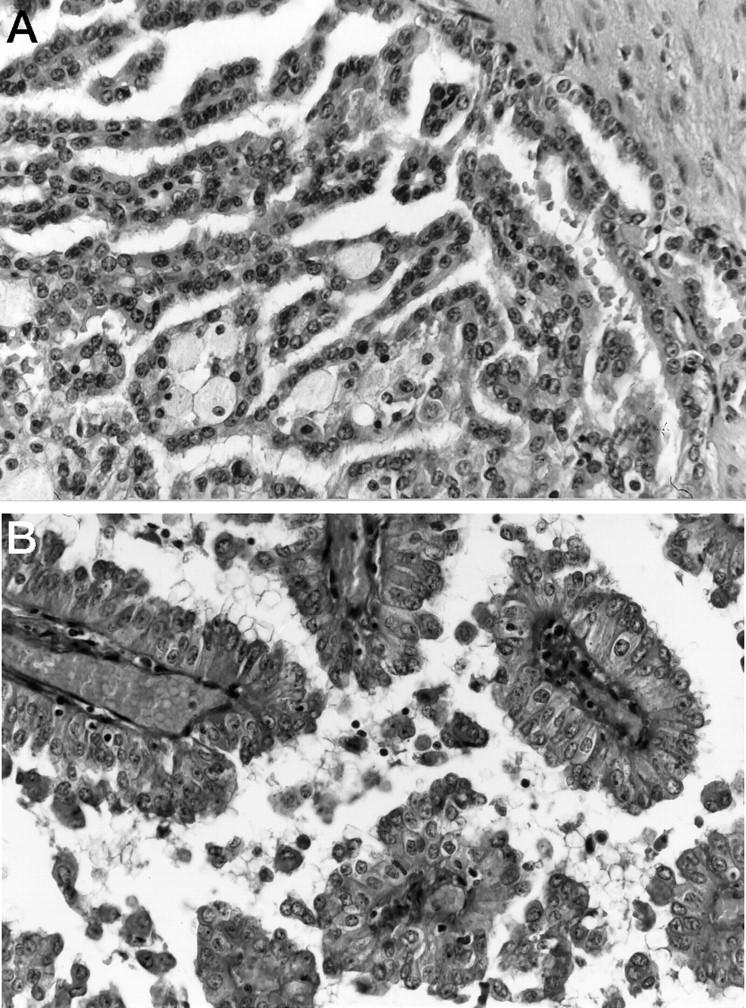
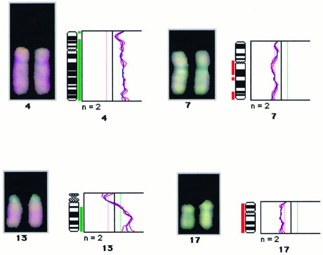
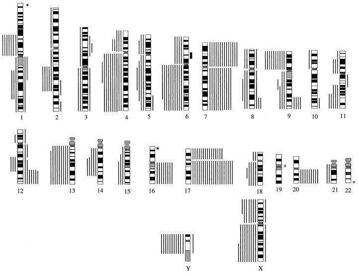
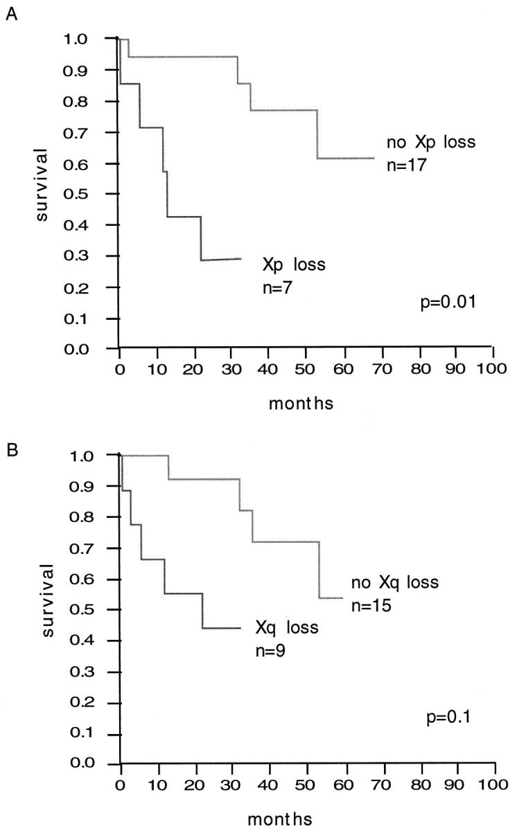
Papillary renal-cell carcinoma (RCC) is a renal carcinoma variant with distinct gross, microscopic, and cytogenetic features. Recently, a type 1 (pale cytoplasm, small-cell) and a type 2 (eosinophilic cytoplasm, large-cell) subtype of papillary RCC have been described. Chromosomal alterations associated with these tumor types were examined in 25 papillary RCCs by comparative genomic hybridization. Relative copy number gains were frequently detected at chromosomes 7p (56%), 7q (44%), 12q (28%), 16q (32%), 17p (56%), 17q (76%), and 20q (32%). Chromosomal regions that were most often lost included 1p (24%), 4q (36%), 6q (40%), 9p (36%), 13q (36%), Xp (28%), Xq (36%), and Y (73%). There were clinical and genetic differences between the subtypes of papillary RCC. Type 2 tumors were of higher nuclear grade (P = 0.0012) and higher stage (P = 0.01) and had a worse prognosis (P = 0.03) than type 1 tumors. The number of DNA gains per tumor, especially gains of 7p and 17p, was significantly higher in type 1 than in type 2 tumors (P < 0.01). These data suggest the existence of two distinct morphological and genetic subgroups of papillary RCC. Losses of chromosome Xp were associated with short patient survival (P < 0.01). Despite the small number of cases, this finding suggests that a gene on chromosome Xp may contribute to papillary RCC progression.
Figures
References
-
- Amin M, Corless C, Renshaaw A, Tickoo S, Kubus J, Schultz D: Papillary (chromophil) renal cell carcinoma: histomorphologic characteristics and evaluation of conventional pathologic prognostic parameters in 62 cases. Am J Surg Pathol 1997, 21:621-635 - PubMed
-
- Mancilla-Jimenez R, Stanley RJ, Blath RA: Papillary renal cell carcinoma. Cancer 1976, 38:2469-2480 - PubMed
-
- Anglard P, Tory K, Brauch H, Weiss GH, Latif F, Merino MJ, Lerman MI, Zbar B, Linehan WM: Molecular analysis of genetic changes in the origin and development of renal cell carcinoma. Cancer Res 1991, 51:1071-1077 - PubMed
-
- Brooks JD, Bova GS, Marshall FF, Isaacs WB: Tumor suppressor gene allelic loss in human renal cancers. J Urol 1993, 150:1278-1283 - PubMed
-
- el Naggar AK, Batsakis JG, Wang G, Lee MS: PCR-based RFLP screening of the commonly deleted 3p loci in renal cortical neoplasms. Diagn Mol Pathol 1993, 2:269-276 - PubMed
Publication types
MeSH terms
LinkOut - more resources
Full Text Sources
Other Literature Sources
Medical
