

Cytoplasmic redistribution of E-cadherin-catenin adhesion complex is associated with down-regulated tyrosine phosphorylation of E-cadherin in human bronchopulmonary carcinomas
- PMID: 9811344
- PMCID: PMC1853397
- DOI: 10.1016/s0002-9440(10)65740-9
Cytoplasmic redistribution of E-cadherin-catenin adhesion complex is associated with down-regulated tyrosine phosphorylation of E-cadherin in human bronchopulmonary carcinomas
Abstract
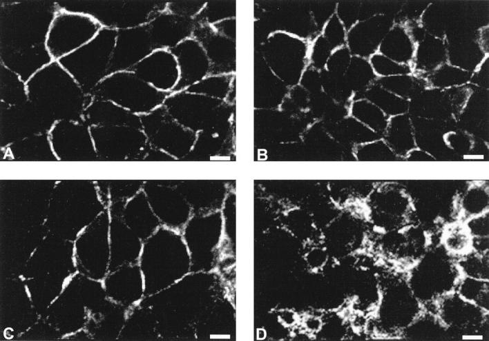
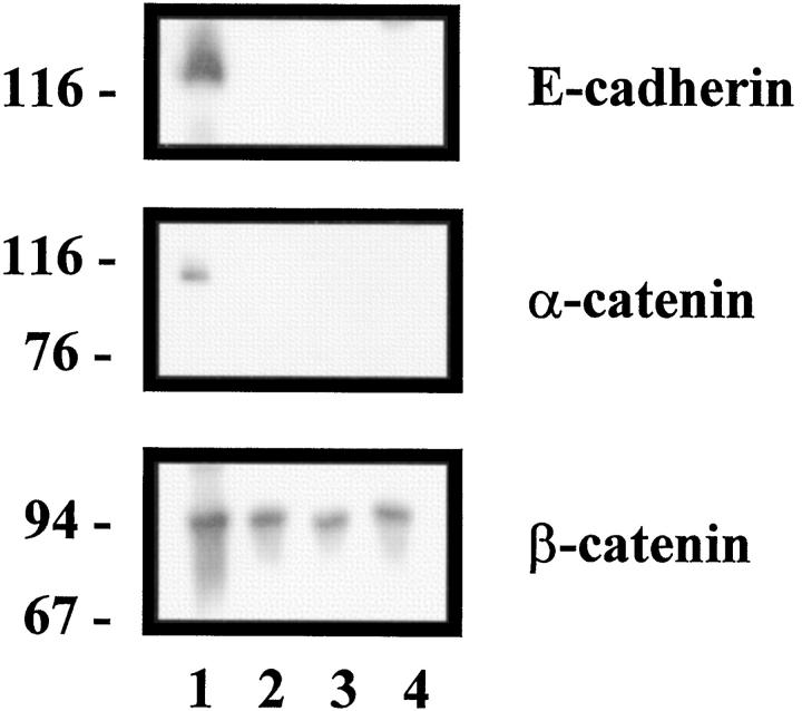
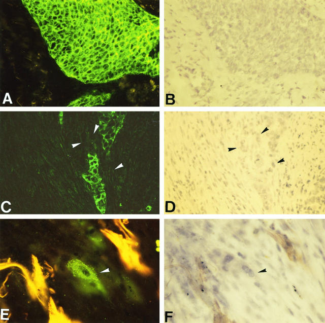
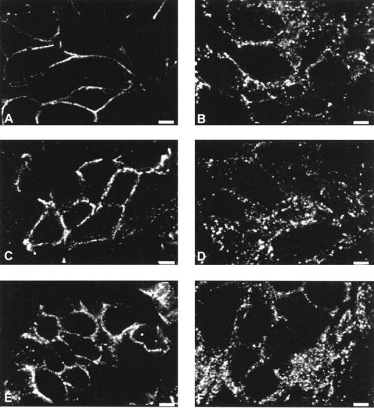
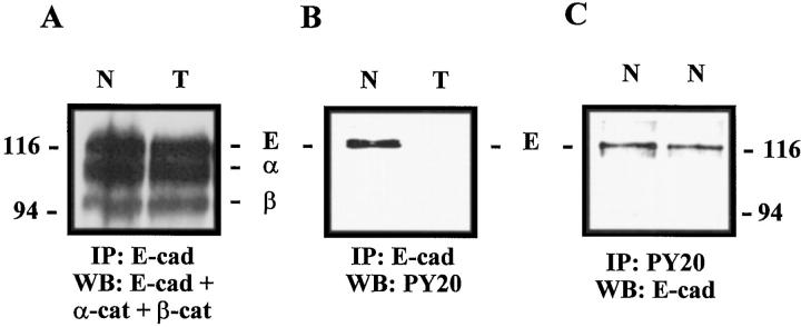
The E-cadherin-catenin complex, by mediating intercellular adhesion, regulates the architectural integrity of epithelia. Down-regulation of its expression is thought to contribute to invasion of carcinoma cells. To investigate the involvement of the E-cadherin-catenin adhesion system in the progression of human bronchopulmonary carcinomas, we compared the immunohistochemical distribution of E-cadherin, alpha-catenin, and beta-catenin in four human bronchial cancer cell lines with different invasive abilities and in 44 primary bronchopulmonary tumors. Although invasive bronchial cell lines did not express E-cadherin and alpha-catenin, complete down-regulation of cadherin-catenin complex expression was a rare event in vivo in bronchopulmonary carcinomas. Nevertheless, a spotty and cytoplasmic pattern of E-cadherin and catenins was observed in 32 primary tumors, only in invasive tumor clusters. Immunoprecipitation experiments showed that this redistribution was not related to a disruption of cadherin-catenin interaction but to down-regulated tyrosine phosphorylation of E-cadherin. We conclude that loss of E-cadherin and/or catenins is not a prominent early event in the invasive progression of human bronchopulmonary carcinomas in vivo. The decreased tyrosine phosphorylation of E-cadherin may reflect a loss of functionality of the complex and implicates a major role in tumor invasion.
Figures
References
-
- Huber O, Bierkamp C, Kemler R: Cadherins and catenins in development. Curr Opin Cell Biol 1996, 8:685-691 - PubMed
-
- Pignatelli M, Vessey CJ: Adhesion molecules: novel molecular tools in tumor pathology. Hum Pathol 1994, 25:849-856 - PubMed
-
- Elangbam CS, Qualls CW, Jr, Dahlgren RR: Cell adhesion molecules: update. Vet Pathol 1997, 34:61-73 - PubMed
-
- Albelda SM: Role of integrins and other cell adhesion molecules in tumor progression and metastasis. Lab Invest 1993, 68:4-17 - PubMed
-
- Munro SB, Blaschuk OW: The structure, function and regulation of cadherins. Brodt P eds. In Cell Adhesion and Invasion in Cancer Metastasis. 1996, :pp 17-34 RG Landes Company, Springer, New York
Publication types
MeSH terms
Substances
LinkOut - more resources
Full Text Sources
Medical
Miscellaneous
