

Pathological findings in human autoimmune lymphoproliferative syndrome
- PMID: 9811346
- PMCID: PMC1853411
- DOI: 10.1016/S0002-9440(10)65742-2
Pathological findings in human autoimmune lymphoproliferative syndrome
Abstract
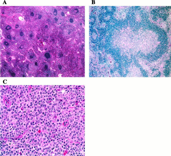
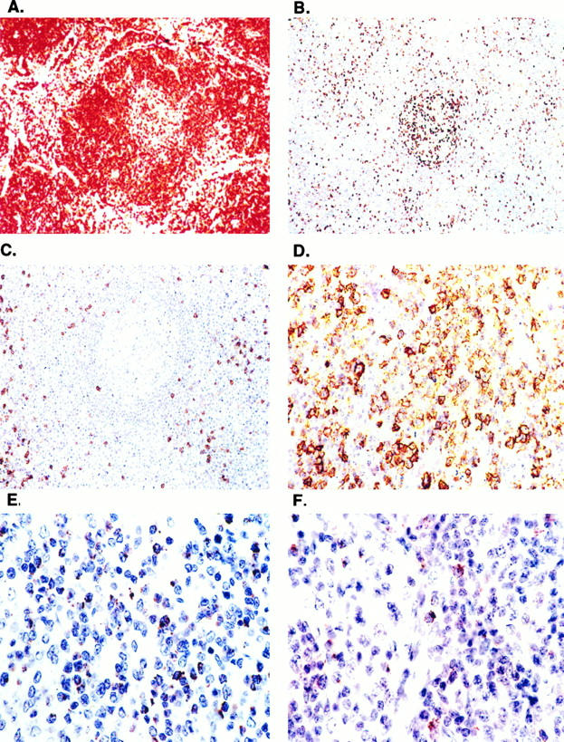
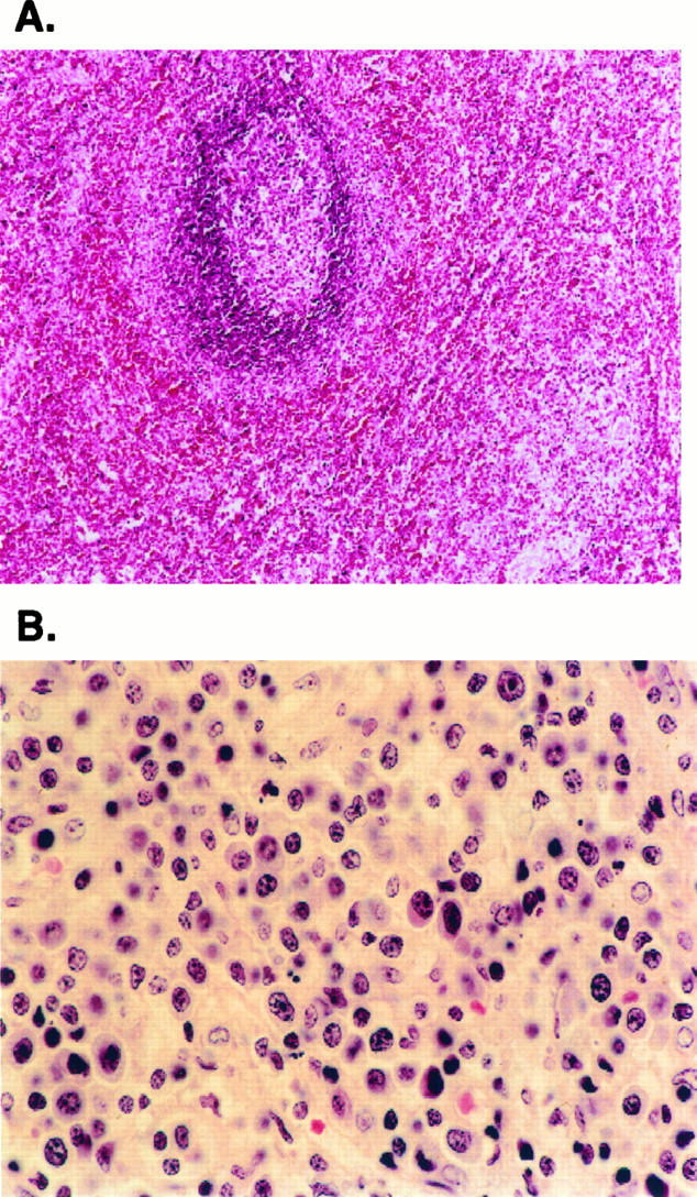
The defects in lymphocyte apoptosis that underlie the autoimmune lymphoproliferative syndrome (ALPS) are usually attributable to inherited mutations of the CD95 (Fas) gene. In this report, we present the histopathological and immunophenotypic features seen in the lymph nodes (n = 16), peripheral blood (n = 10), bone marrow (n = 2), spleen (n = 3), and liver (n = 2) from 10 patients with ALPS. Lymph nodes showed marked paracortical hyperplasia. Interfollicular areas were expanded and populated by T cell receptor-alphabeta CD3+ CD4-CD8- (double-negative, DN) T cells that were negative for CD45RO. CD45RA+ T cells were increased in all cases studied. The paracortical infiltrate was a result of both reduced apoptosis and increased proliferation, as measured by in situ detection of DNA fragmentation and staining with MIB-1, respectively. The paracortical proliferation may be extensive enough to suggest a diagnosis of malignant lymphoma. Many of the paracortical lymphocytes expressed markers associated with cytotoxicity, such as perforin, TIA-1, and CD57. CD25 was negative. In addition, most lymph nodes exhibited florid follicular hyperplasia, often with focal progressive transformation of germinal centers; in some cases, follicular involution was seen. A polyclonal plasmacytosis also was present. The spleens were markedly enlarged, more than 10 times normal size. There was expansion of both white pulp and red pulp, with increased DN T cells. DN T cells also were observed in liver biopsies exhibiting portal triaditis. In the peripheral blood, the T cells showed increased expression of HLA-DR and CD57 but not CD25. CD45RA+ T cells were increased in the four cases studied. Polyclonal B cell lymphocytosis with expansion of CD5+ B cells was a characteristic finding. Taken together, the histopathological and immunophenotypic findings, particularly in lymph nodes and peripheral blood, are sufficiently distinctive to suggest a diagnosis of ALPS. Of note, two affected family members of one proband developed lymphoma (T-cell-rich B-cell lymphoma and nodular lymphocyte predominance Hodgkin's disease, respectively).
Figures
References
-
- Watanabe-Fukunaga R, Brannan PE, Copeland NG, Jenkins N, Nagata S: Lymphoproliferation disorder in mice explained by defects in Fas antigen that mediates apoptosis. Nature 1992, 356:314-317 - PubMed
-
- Takahashi T, Tanaka M, Rannan CI, Jenkins NA, Copeland NG, Suda T, Nagata S: Generalized lymphoproliferative disease in mice, caused by a point mutation in the Fas ligand. Cell 1994, 76:969-977 - PubMed
-
- Lynch DH, Watson ML, Alderson MR, Baum PR, Miller RE, Tough T, Gibson M, Davis-Smith T, Smith CA, Hunter K, Bhat D, Din W, Goodwin RG, Seldin MF: The mouse Fas-ligand gene is mutated in gld mice and is part of a TNF family gene cluster. Immunity 1994, 1:131-140 - PubMed
-
- Fisher GH, Rosenberg FJ, Straus SE, Dale JK, Middleton LA, Lin AY, Strober W, Lenardo MJ, Puck JM: Dominant interfering Fas gene mutations impair apoptosis in a human autoimmune lymphoproliferative syndrome. Cell 1995, 81:935-946 - PubMed
Publication types
MeSH terms
Substances
LinkOut - more resources
Full Text Sources
Medical
Research Materials
Miscellaneous
