. 1998 Nov;180(22):5947-53.
doi: 10.1128/JB.180.22.5947-5953.1998.

Autolysis of Lactococcus lactis is influenced by proteolysis

Affiliations

Autolysis of Lactococcus lactis is influenced by proteolysis

G Buist et al. J Bacteriol. 1998 Nov.

Abstract

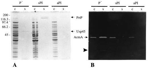
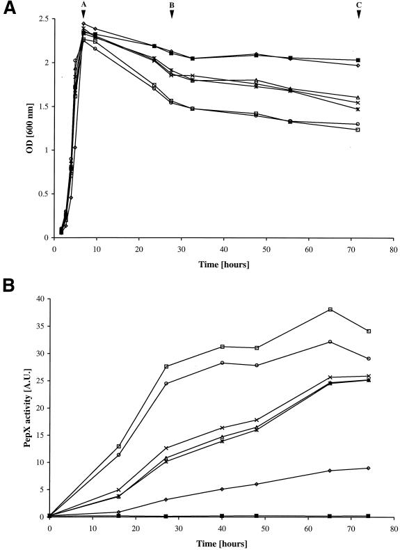
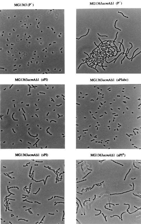
The autolysin AcmA of Lactococcus lactis was shown to be degraded by the extracellular lactococcal proteinase PrtP. Autolysis, as evidenced by reduction in optical density of a stationary-phase culture and concomitant release of intracellular proteins, was greatly reduced when L. lactis MG1363 cells expressed the cell wall-anchored lactococcal proteinase PrtP of the PI-type caseinolytic specificity (PI). On the other hand, lactococcal strains that did not produce the proteinase showed a high level of autolysis, which was also observed when the cells produced the secreted form of PI or a cell wall-anchored proteinase with PIII-type specificity. Autolysis was also increased when MG1363 expressed the cell wall-anchored hybrid PI/PIII-type proteinase PIac. Zymographic analysis of AcmA activity during stationary phase showed that AcmA was quickly degraded by PI and much more slowly by PrtP proteinases with PIII-type and intermediate specificities. Autolysis of L. lactis by AcmA was influenced by the specificity, amount, and location of the lactococcal proteinase. No autolysis was observed when the various proteinases were expressed in an L. lactis acmA deletion mutant, indicating that PrtP itself did not cause lysis of cells. The chain length of a strain was significantly shortened when the strain expressed a cell wall-anchored active proteinase.


Figures

FIG. 1
FIG. 1
(A) Protein profiles of cell (c) and supernatant (s) fractions of whey-grown L. lactis MG1363 containing pGK13 (P), pGKV500 (sPI), or pGKV552 (aPI) in an SDS–12.5% polyacrylamide gel stained with Coomassie brilliant blue. PrtP, lactococcal proteinase; Usp45, secreted protein of unknown function (28). (B) Analysis of AcmA activity in the samples used for panel A by renaturing SDS-PAGE (12.5% polyacrylamide gel) in the presence of 0.15% M. lysodeikticus autoclaved cells. AcmA, N-acetylmuramidase of L. lactis; arrowhead, major degradation product of AcmA (5). Molecular masses (in kilodaltons) of standard proteins for both gels are shown on the left. Five microliters of each sample was loaded onto the gels.
FIG. 2
FIG. 2
(A) Growth and lysis by optical density (OD) measurement at 600 nm of cultures of L. lactis MG1363 containing pGK13 (P; ×), pGKV500 (sPI; ∗), pGKV1552 (aPI*; ○), pGKV552 (aPI; ◊), pGKV552abc (aPIabc; ▵), or pGKV552ac (aPIac; □). L. lactis MG1363acmAΔ1 containing either of these plasmids exhibited very similar growth curves of which only one, MG1363acmAΔ1(pGK13), is shown (■). The strains were examined during 3 days of incubation at 30°C. Lettered arrowheads indicate the time points at which samples were taken from the cultures for analysis of AcmA activity (Fig. 3) and protein release (Fig. 2B and data not shown). (B) Analysis of PepX activity (in arbitrary units [A.U.]) in the culture supernatants of the strains presented in panel A. Symbols are as defined for panel A.
FIG. 3
FIG. 3
Zymographic analysis of AcmA activity. Each lane contains 10 μl of the supernatant of an MG1363 culture in M17 producing the indicated proteinase sampled at the time points A, B, and C shown in Fig. 2A. Molecular masses (in kilodaltons) of standard proteins are shown on the left.
FIG. 4
FIG. 4
Light microscopic analysis of the chain lengths in overnight M17 cultures of the indicated strains. Magnification, ×1,000.

References

    1. Ayusawa D, Yoneda Y, Yamane K, Maruo B. Pleiotropic phenomena in autolytic enzyme(s) content, flagellation, and simultaneous hyperproduction of extracellular α-amylase and protease in a Bacillus subtilis mutant. J Bacteriol. 1975;124:459–469. - PMC - PubMed
    1. Béliveau C, Potvin C, Trudel J, Asselin A, Bellemare G. Cloning, sequencing, and expression in Escherichia coli of a Streptococcus faecalis autolysin. J Bacteriol. 1991;173:5619–5623. - PMC - PubMed
    1. Buist G. AcmA of Lactococcus lactis, a cell-binding muramidase. Ph.D. thesis. Groningen, The Netherlands: University of Groningen; 1997.
    1. Buist G, Karsens H, Nauta A, van Sinderen D, Venema G, Kok J. Autolysis of Lactococcus lactis caused by induced overproduction of its major autolysin, AcmA. Appl Environ Microbiol. 1997;63:2722–2728. - PMC - PubMed
    1. Buist G, Kok J, Leenhouts K J, Dabrowska M, Venema G, Haandrikman A J. Molecular cloning and nucleotide sequence of the gene encoding the major peptidoglycan hydrolase of Lactococcus lactis, a muramidase needed for cell separation. J Bacteriol. 1995;177:1554–1563. - PMC - PubMed

Publication types

MeSH terms