. 1998 Dec;72(12):9788-94.
doi: 10.1128/JVI.72.12.9788-9794.1998.

Neutralizing monoclonal antibodies block human immunodeficiency virus type 1 infection of dendritic cells and transmission to T cells

Affiliations

Neutralizing monoclonal antibodies block human immunodeficiency virus type 1 infection of dendritic cells and transmission to T cells

S S Frankel et al. J Virol. 1998 Dec.

Abstract

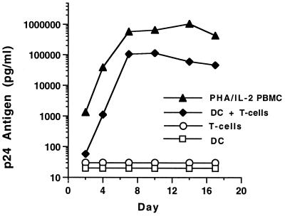
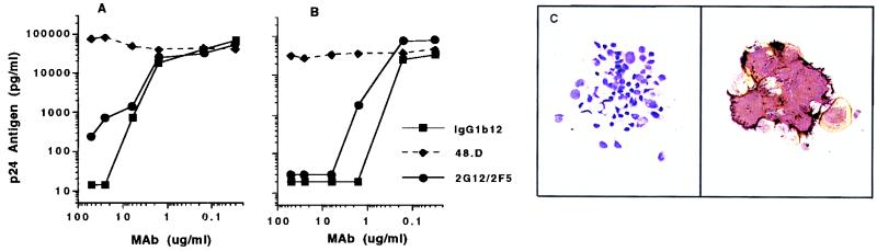
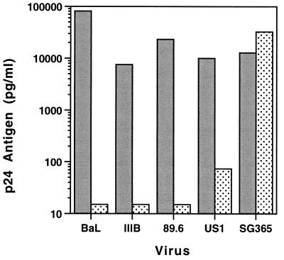
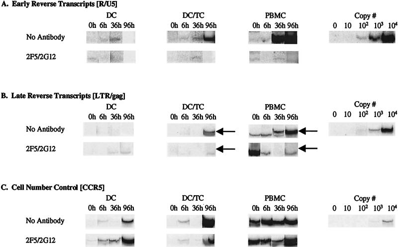
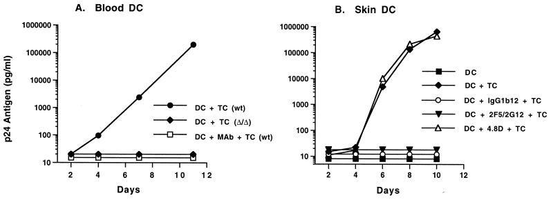
Prevention of the initial infection of mucosal dendritic cells (DC) and interruption of the subsequent transmission of HIV-1 from DC to T cells are likely to be important attributes of an effective human immunodeficiency virus type 1 (HIV-1) vaccine. While anti-HIV-1 neutralizing antibodies have been difficult to elicit by immunization, there are several human monoclonal antibodies (MAbs) that effectively neutralize virus infection of activated T cells. We investigated the ability of three well-characterized neutralizing MAbs (IgG1b12, 2F5, and 2G12) to block HIV-1 infection of human DC. DC were generated from CD14(+) blood cells or obtained from cadaveric human skin. The MAbs prevented viral entry into purified DC and the ensuing productive infection in DC/T-cell cultures. When DC were first pulsed with HIV-1, MAbs blocked the subsequent transmission to unstimulated CD3(+) T cells. Thus, neutralizing antibodies can block HIV-1 infection of DC and the cell-to-cell transmission of virus from infected DC to T cells. These data suggest that neutralizing antibodies could interrupt the initial events associated with mucosal transmission and regional spread of HIV-1.


Figures

FIG. 1
FIG. 1
Infection kinetics of purified DC cultured in the presence and absence of T cells. The DC were exposed to HIV-1 BaL (500 TCID50/105 DC) overnight at 37°C, thoroughly washed to remove free virus, and cultured alone or with positively selected CD3+ T cells. PHA/IL-2-stimulated PBMC and unstimulated CD3+ cells were infected in a similar manner. No p24 antigen expression was detected in purified DC or CD3+ T cells alone. Robust infection, close to that seen with activated PBMC, was detected when T cells were added to infected DC. During infection, DC and CD3+ cells were cultured in medium without exogenous cytokines. PBMC were maintained in culture medium supplemented with 20 U of IL-2 per ml.
FIG. 2
FIG. 2
(A and B) Antibody-mediated neutralization of HIV-1 BaL infection of PHA/IL-2 stimulated PBMC (A) and DC (B), with CD3+ cells added on day 1 after exposure of DC to HIV-1. MAb IgG1b12, or the combination 2F5/2G12, was preincubated with virus at a starting concentration (for each MAb) of 50 μg/ml prior to addition of PBMC or DC. Extracellular p24 antigen was measured in the early viral growth phase for PBMC (day 4) and DC/T cells (day 7). Identical concentrations of the nonneutralizing MAb 4.8D did not inhibit viral infection of PBMC or DC targets. (C) Immunocytochemical staining of DC/T-cell cytospins for p24 antigen expression (brown) and HLA-DR (purple). The right-hand panel shows a p24-positive syncytium on day 7 after DC exposure to BaL (magnification, ×200). No HIV-1-infected cells were seen when BaL was preincubated with 25 μg of MAb IgG1b12 per ml (left, p24 antigen stain only; ×160).
FIG. 3
FIG. 3
Neutralization of four clade B HIV-1 isolates by the combination 2F5/2G12 (each at 25 μg/ml). Target cells were DC with T cells added back as in Fig. 1. Stippled bars indicate preincubation of DC with MAbs; gray bars show virus growth without antibody. The clade C isolate (SG365) was not neutralized by 2F5/2G12.
FIG. 4
FIG. 4
Neutralizing MAbs block virus entry into DC. Purified blood DC were exposed to a DNase-treated, cell-free virus stock (HIV-1 BaL; MOI = 0.01) in the presence or absence of MAb combination 2F5/2G12 (each at 25 μg/ml) and subsequently incubated alone (DC) or in the presence of purified CD3+ T cells (DC/TC). Parallel control infections of PHA/IL-2-stimulated PBMC were also conducted. Cell lysates were prepared at the times indicated postinfection and amplified with 32P-labeled primers specific for early and late HIV-1 reverse transcripts and for CCR5. Parallel amplification of known numbers of ACH-2 cells (which contain a single integrated HIV-1 provirus per cell) were performed with each primer set to provide a copy number quantitation curve. (A) Early reverse transcripts (RU5 long terminal repeat [LTR] region sequences). (B) Late reverse transcripts (LTR/gag). Arrows indicate the positions of specific signals; the remaining visible bands are nonspecific (i.e., no late reverse transcripts were seen in PBMC until the 36-h time point). (C) CCR5 gene amplification (cell number control).
FIG. 5
FIG. 5
Neutralizing MAbs prevent the transmission of HIV-1 from infected DC to T cells. Purified blood (A) or skin (B) DC were exposed to HIV-1 BaL for 90 min and washed to remove free virus. MAbs were added 24 h later. After a 30-min incubation, CD3+ T cells were added to the culture. (A) MAb IgGb12 was used at 25 μg/ml. As a control, CD3+ cells from a known homozygous CCR5 (Δ/Δ) donor were also used. Transmission of infection from DC to T cells was dependent on CCR5 and could be blocked by neutralizing MAbs. wt, T cells wild type for the CCR5 gene. (B) MAbs were used at a concentration of 25 μg/ml.

References

    1. Ayehunie S, Garcia-Zepeda E A, Hoxie J A, Horuk R, Kupper T S, Luster A D, Ruprecht R M. Human immunodeficiency virus-1 entry into purified blood dendritic cells through CC and CXC chemokine coreceptors. Blood. 1997;90:1379–1386. - PubMed
    1. Ayehunie S, Groves R W, Bruzzese A-M, Ruprecht R M, Kupper T S, Langhoff E. Acutely infected Langerhans cells are more efficient than T cells in disseminating HIV type 1 to activated T cells following a short cell-cell contact. AIDS Res Hum Retroviruses. 1995;11:877–884. - PubMed
    1. Banchereau J, Steinman R M. Dendritic cells and the control of immunity. Nature. 1998;392:245–252. - PubMed
    1. Bender A, Sapp M, Schuler G, Steinman R M, Bhardwaj N. Improved methods for the generation of dendritic cells from nonproliferating progenitors in human blood. J Immunol Methods. 1996;196:121–135. - PubMed
    1. Blauvelt A, Asada H, Saville M W, Klaus-Kovtun V, Altman D J, Yarchoan R, Katz S I. Productive infection of dendritic cells by HIV-1 and their ability to capture virus are mediated through separate pathways. J Clin Investig. 1997;100:2043–2053. - PMC - PubMed

Publication types