. 1998 Dec;72(12):9795-805.
doi: 10.1128/JVI.72.12.9795-9805.1998.

Successful readministration of adeno-associated virus vectors to the mouse lung requires transient immunosuppression during the initial exposure

Affiliations

Successful readministration of adeno-associated virus vectors to the mouse lung requires transient immunosuppression during the initial exposure

C L Halbert et al. J Virol. 1998 Dec.

Abstract

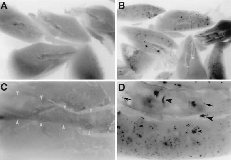
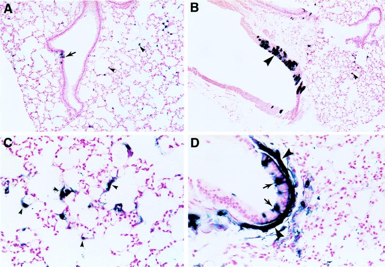
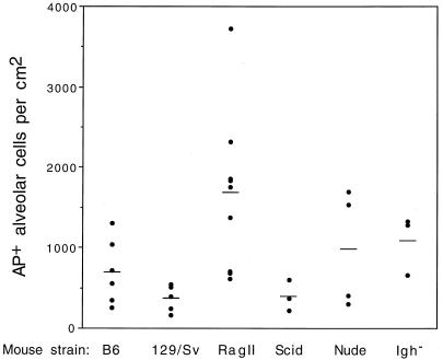
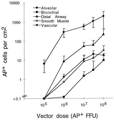
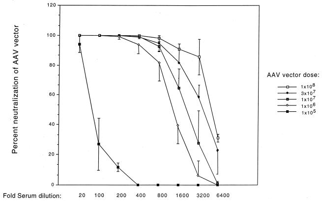
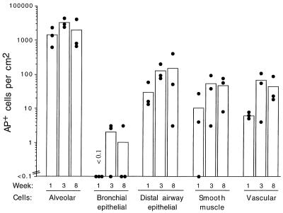
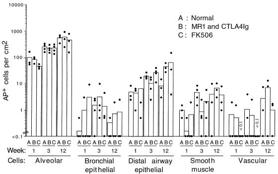
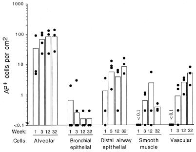
The airway is an important target for gene transfer to treat cystic fibrosis and other diseases that affect the lung. We previously found that marker gene expression did not persist in the bronchial epithelium following adeno-associated virus (AAV) vector administration to the rabbit lung. In an attempt to promote continued expression, we tested repeat vector administration, but no additional transduction was observed, and the block to transduction correlated with the appearance of neutralizing antibodies to the viral capsid. Here we show that mice exhibit a similar response but that treatment with anti-CD40 ligand antibody (MR1) and a soluble CTLA4-immunoglobulin fusion protein (CTLA4Ig) at the time of primary AAV vector exposure allowed successful repeat transduction and prevented production of neutralizing antibodies. We also tested the possibility that an immune response caused the loss of marker-positive cells in the epithelial population in rabbits by evaluating AAV vector expression in immunocompetent and immunodeficient mice. In contrast to results in rabbits, marker protein expression persisted in the lung in both groups of mice. AAV vector transduction occurred in alveolar cells, airway epithelial cells, and smooth muscle cells, and vector expression persisted for at least 8 months. Although data on persistence of AAV vector expression in the human lung are not available, it is likely that repeat transduction will be necessary either due to loss of expression or to the need for repeat administration to deliver effective amounts of AAV vectors. Results presented here indicate that transient immunosuppression will allow such repeat vector treatment of the lung.


Figures

FIG. 1
FIG. 1
AAV vector transduction in the mouse lung. Mice were given saline (A and C) or 107 AP+ FFU of AAV-AP (made by using the AAV/Ad packaging plasmid) (B and D) by intratracheal inoculation. The lungs were excised and stained for AP expression 21 days after inoculation. (A and C) Saline-treated mouse lungs do not exhibit any AP+ cells. The bronchus is outlined by gray arrowheads in panel C. (B and D) AAV-AP-treated lungs show AP staining in epithelial cells (arrows) and smooth muscle cells (large arrowheads) of bronchial airways and in parenchymal cells (small arrowheads) of the lung. Original magnifications, ×8 (A and B) and ×32 (C and D).
FIG. 2
FIG. 2
Histologic analysis of AAV vector-treated mouse lungs. Mice were given AAV-AP and were treated as described in the legend to Fig. 1. AP staining was performed 21 days after vector exposure. AP+ epithelial cells in distal airways are indicated in panels A and D by arrows. AP+ alveolar cells are designated by a small arrowheads in panes A, B, and C. AP+ smooth muscle cells were found beneath the airway epithelium (large arrowheads in panel D) and in vascular walls (large arrowhead in panel B). Original magnifications, ×100 (A and B) and ×400 (C and D).
FIG. 3
FIG. 3
Transduction by AAV vector in immunocompetent and immunodeficient strains of mice. Mice were treated as described in the legend to Fig. 1. AP staining was performed 21 days after vector exposure. Transduction efficiencies in individual animals (solid circles) and mean values (bars) are shown.
FIG. 4
FIG. 4
Relationship of AAV vector dose to transduction efficiency in different cell populations of the mouse lung. B6 mice were given the indicated doses of the AAV-AP vector (made by using the pMTrepCMVcap packaging plasmid) by nasal aspiration. The lungs were excised 21 days after vector exposure and stained for AP expression. Arithmetic mean values ± standard deviations for the transduction rates are shown (n = 4 for each vector dose tested). Transduction rates for all cell types in animals receiving vector doses of ≥107 AP+ FFU were significantly different (P ≤ 0.05) from background.
FIG. 5
FIG. 5
Relationship of AAV vector dose to generation of neutralizing antibodies against the AAV vector. Mice were treated as described in the legend to Fig. 4, and sera were obtained from animals 21 days after exposure to the AAV-AP vector. Percent neutralization of the AAV-βgal vector is shown. Duplicate assays were done for each serum dilution for all animals (n = 4 animals for each vector dose). Mean values ± standard deviations for percent neutralization at each serum dilution are shown.
FIG. 6
FIG. 6
Persistence of AAV vector expression in the different cell populations of the RagII mouse lung. Mice were given 107 AP+ FFU of AAV-AP (made by using the AAV/Ad packaging plasmid) by intratracheal inoculation, groups of mice were sacrificed at the indicated times after exposure, and the lungs were stained for AP. Three animals in each group were analyzed. Means (bars) and individual values (solid circles) are shown.
FIG. 7
FIG. 7
Persistence of AAV vector expression in the different cell populations of the B6 mouse lung. Mice were given 107 AP+ FFU of AAV-AP (made by using the AAV/Ad packaging plasmid) by intratracheal inoculation. Animals were either untreated (A), treated with MR1 and CTLA4Ig (B), or treated with FK506 (C). Three animals were analyzed for weeks 1 and 12, and four animals were analyzed for week 3. Means (bars) and individual values (solid circles) are shown.
FIG. 8
FIG. 8
Persistence of AAV vector expression in the different cell populations of BALB/c mice. Mice were treated as described in the legend to Fig. 6. Three animals were analyzed for weeks 1, 12, and 32, and four animals were analyzed for week 3. Means (bars) and individual values (solid circles) are shown.

References

    1. Alexander I E, Russell D W, Spence A M, Miller A D. Effects of gamma irradiation on the transduction of dividing and nondividing cells in brain and muscle of rats by adeno-associated virus vectors. Hum Gene Ther. 1996;7:841–850. - PubMed
    1. Allen J M, Debelak D J, Reynolds T C, Miller A D. Identification and elimination of replication-competent adeno-associated virus (AAV) that can arise by nonhomologous recombination during AAV vector production. J Virol. 1997;71:6816–6822. - PMC - PubMed
    1. Ayers M M, Jeffery P K. Proliferation and differentiation in mammalian airway epithelium. Eur Respir J. 1988;1:58–80. - PubMed
    1. Bar D, Tubb J, Ferguson D, Scaria A, Lieber A, Wilson C, Perkins J, Kay M A. Strain related variation in adenovirally mediated transgene expression from mouse hepatocytes in vivo: comparisons between immunocompetent and immunodeficient inbred strains. Gene Ther. 1995;2:151–155. - PubMed
    1. Bierer B E. Mechanisms of action of immunosuppressive agents: cyclosporin A, FK506, and rapamycin. Proc Assoc Am Physicians. 1995;107:28–40. - PubMed

Publication types

MeSH terms