. 1998 Dec;72(12):9906-17.
doi: 10.1128/JVI.72.12.9906-9917.1998.

Genetic analysis of the rat leukemia virus: influence of viral sequences in transduction of the c-ras proto-oncogene and expression of its transforming activity

Affiliations

Genetic analysis of the rat leukemia virus: influence of viral sequences in transduction of the c-ras proto-oncogene and expression of its transforming activity

S Y Lee et al. J Virol. 1998 Dec.

Abstract

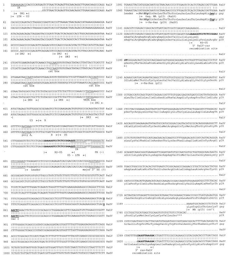
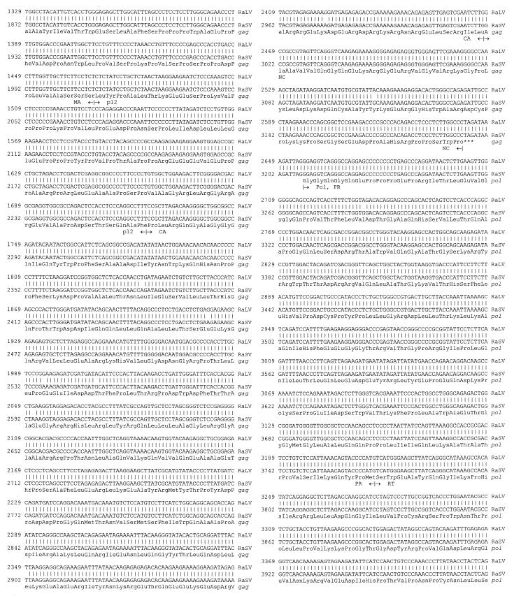
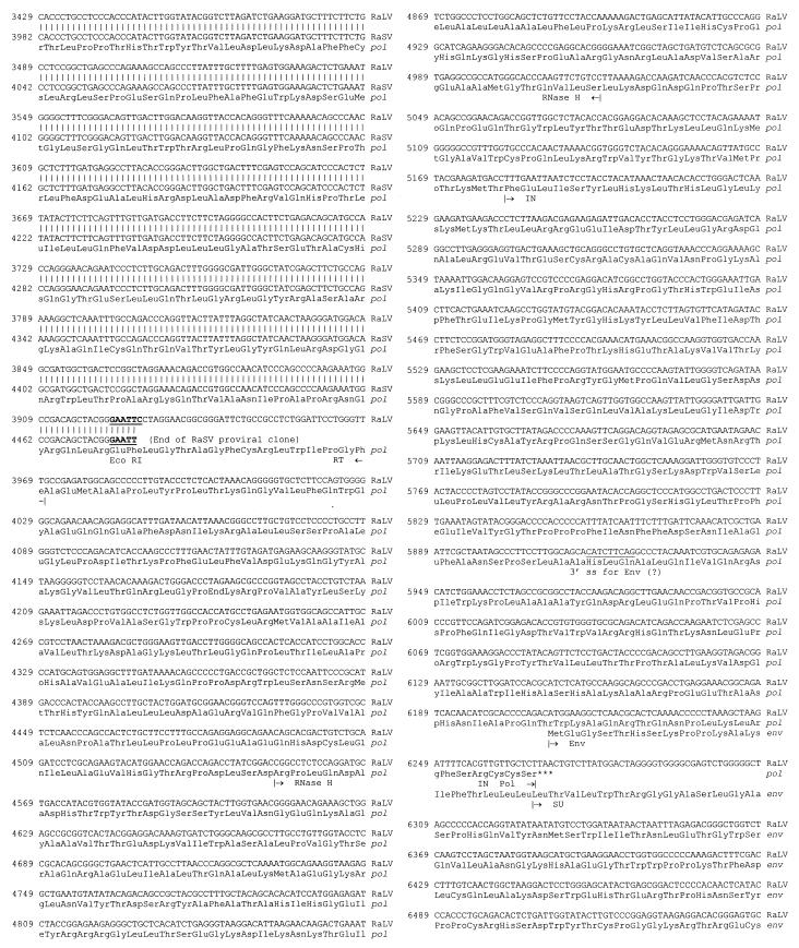
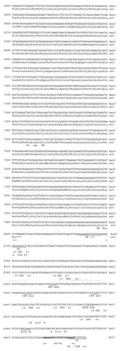
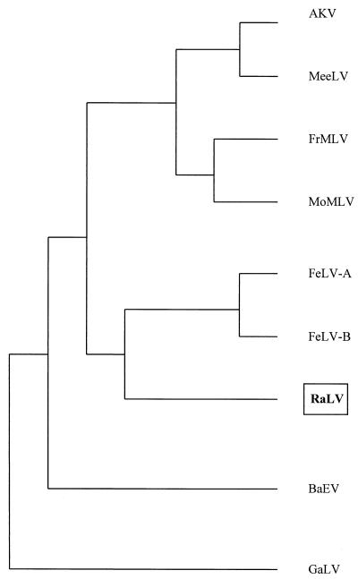
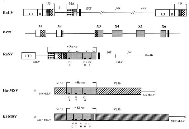
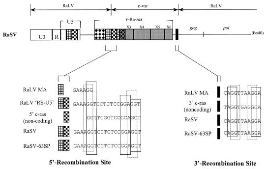
The rat leukemia virus (RaLV) is an endogenous retrovirus that is spontaneously released by Sprague-Dawley rat embryo cells. The overall structure of the RaLV genome resembles that of other simple, replication-competent retroviruses, but the sequence of the long terminal repeats (LTR) is unique and unrelated to the known retroviruses. Phylogenetically, the RaLV genome appears to be more closely related to the feline leukemia virus group of retroviruses than to the murine leukemia virus group. A remarkable feature of RaLV is that it is capable of transducing a ras proto-oncogene from rat tumor cells in the form of an acutely transforming virus, designated the Rasheed strain of the rat sarcoma virus (RaSV). With the exception of the c-ras sequence, the genomes of both RaLV and RaSV are collinear. The RaSV-encoded oncogene v-Ra-ras expresses a fusion protein with a molecular mass of 29 kDa, and it exhibits a unique structure that has not been described previously for any known virus. The 5' end of this gene is derived from sequences encoding RaLV matrix followed by 20 bp derived from the U5 region of the RaLV LTR (RS-U5 element) which is joined at its 3' end to sequences derived from all six (coding and noncoding) exons of the c-ras proto-oncogene at the 3' end. This recombinational event represents a novel mechanism among the acutely transforming viruses that have been studied.


Figures

FIG. 1
FIG. 1
Nucleotide sequence alignment of RaLV and RaSV. The complete nucleotide sequence of the RaLV genome and the 5′ end of the RaSV genome is shown together with the deduced amino acid sequences of the RaLV Gag, Pol, and Env proteins and the RaSV p29 oncoprotein (v-Ra-Ras). The RaSV sequence was aligned to RaLV by the Gap (GCG) program. Dots represent gaps which were inserted into both sequences to maintain the alignment, and vertical lines between the sequences represent agreement between the two genomes. The cloned RaLV genome contained 8,107 bp and one LTR. To properly display the alignment between the RaLV and RaSV genomes, a deduced 8,695-bp clone RaLV provirus with two LTRs is displayed. The boundaries of various regions of viral genomes are indicated by arrows. The nucleotides comprising the 5′ and 3′ RaLV-ras recombination sites are depicted in boldface, italic letters, and the sequence of the RS-U5 element in the LTR is in boldface type. Other notable LTR elements that are demarcated include positions of the CCAAT and TATAA promoter motifs located within the U3 region, the polyadenylation signal sequence within the R region, direct repeats (DR1 to DR4) and the flanking indirect repeats (IR). The primer binding sites for both positive- and negative-strand DNA synthesis are marked as are putative major 5′ and 3′ splice sites for env mRNA expression. The SacI and EcoRI restriction endonuclease sites were utilized to initially clone the RaLV and RaSV genomes, respectively. Labeled in boldface letters is the myristoylation signal sequence at the second amino acid residue (Gly) of the p15gag MA and p29 v-Ra-Ras proteins. The single-amino-acid-residue differences between the amino termini of these two proteins are underscored and in boldface, italic letters.
FIG. 1
FIG. 1
Nucleotide sequence alignment of RaLV and RaSV. The complete nucleotide sequence of the RaLV genome and the 5′ end of the RaSV genome is shown together with the deduced amino acid sequences of the RaLV Gag, Pol, and Env proteins and the RaSV p29 oncoprotein (v-Ra-Ras). The RaSV sequence was aligned to RaLV by the Gap (GCG) program. Dots represent gaps which were inserted into both sequences to maintain the alignment, and vertical lines between the sequences represent agreement between the two genomes. The cloned RaLV genome contained 8,107 bp and one LTR. To properly display the alignment between the RaLV and RaSV genomes, a deduced 8,695-bp clone RaLV provirus with two LTRs is displayed. The boundaries of various regions of viral genomes are indicated by arrows. The nucleotides comprising the 5′ and 3′ RaLV-ras recombination sites are depicted in boldface, italic letters, and the sequence of the RS-U5 element in the LTR is in boldface type. Other notable LTR elements that are demarcated include positions of the CCAAT and TATAA promoter motifs located within the U3 region, the polyadenylation signal sequence within the R region, direct repeats (DR1 to DR4) and the flanking indirect repeats (IR). The primer binding sites for both positive- and negative-strand DNA synthesis are marked as are putative major 5′ and 3′ splice sites for env mRNA expression. The SacI and EcoRI restriction endonuclease sites were utilized to initially clone the RaLV and RaSV genomes, respectively. Labeled in boldface letters is the myristoylation signal sequence at the second amino acid residue (Gly) of the p15gag MA and p29 v-Ra-Ras proteins. The single-amino-acid-residue differences between the amino termini of these two proteins are underscored and in boldface, italic letters.
FIG. 2
FIG. 2
Phylogenetic tree showing relationship of RaLV to other mammalian retroviruses. The deduced amino acid sequences of the Gag, Pol, and Env proteins of RaLV were separately compared by using the program GrowTree (GCG) to their counterparts of other mammalian leukemia viruses found in the protein sequence databases. The neighbor-joining method was used to cluster the sequences in a pairwise fashion prior to construction of the tree (49). The patterns of all three phylogenetic trees were identical. This diagram shows the evolutionary relationships of complete Gag polyproteins. Viruses used in the analysis included murine leukemia virus from the AKR mouse strain (AKV), baboon endogenous virus (BaEV), FeLV types A and B (FeLV-A and -B, respectively), Friend murine leukemia virus (FrMLV), gibbon ape leukemia virus (GaLV), murine (mouse) ecotropic endogenous leukemia virus (MeeLV), MoMLV, and RaLV.
FIG. 3
FIG. 3
Diagrammatic representation of RaLV and c-ras sequences participating in the formation of RaSV (not drawn to scale). Patterned boxes represent specific nucleic acid sequence domains from different regions of the RaLV genome or the c-ras proto-oncogene; boxes with the same design or shading represent similar or identical nucleic acid sequences. The top line shows the RaLV genome with its terminally redundant LTRs and the relative positions of the gag, pol, and env genes and the leader sequence (L). The second line represents the structure of the wild-type c-ras gene; only four exons encode the p21 c-Ras protein (exons 3, 4, 5, and 6). Exons 1 and 2 are noncoding in all ras-containing viruses except RaSV, and the start codon for the p21 protein is located within exon 3. The third line is a graphical representation of the RaSV genome showing the relative positions of sequences derived from either RaLV or c-ras. The majority of the RaSV genome is derived from RaLV; however, the v-Ra-ras oncogene is composed of three domains, two of which are derived from RaLV and the third of which is derived from exons 1 to 6 of the c-ras proto-oncogene. The first 92 bp of the RaLV MA gene comprised the first domain, the second domain consisted of a 20-bp element (RS-U5) derived from the 3′ end of the U5 region of the RaLV LTR, and the 3′-most 638 bp from the c-ras proto-oncogene formed the third. The black boxes in the RaLV, c-ras, and RaSV diagrams represent the positions of the 3′ recombination sequence believed to participate in the formation of RaSV. The fourth and fifth lines are schematic diagrams showing the cellular and viral origins of various regions of the Ha-MSV and Ki-MSV genomes surrounding the v-ras oncogenes incorporated by the respective MuLV strains. These two oncogenes are found to be bracketed by sequences derived from rat VL30 elements. Solid circles within the exons of the v-ras oncogenes represent changes in the amino acid residues of the three sarcoma viruses relative to the sequence of the cellular c-Ras protein.
FIG. 4
FIG. 4
Proposed RaLV and c-ras nucleotide sequences that appear to participate in the formation of the RaSV genome. The top line is a schematic representation depicting the different regions of the RaSV genome. Patterned boxes represent specific nucleic acid sequence domains from different regions of the RaLV genome or the c-ras proto-oncogene; as in Fig. 3, boxes with the same design or shading represent similar or identical nucleic acid sequences. RaSV-63SP is a second RaSV isolate recovered independently in Japan by in vitro cocultivation of RaLV-productive SD1-T cells with a cell line established from a rat mammary tumor induced by dimethylbenzanthracene (27).

References

    1. Altschul S F, Madden T L, Schaffer A A, Zhang J, Zhang Z, Miller W, Lipman D J. Gapped BLAST and PSI-BLAST: a new generation of protein database search programs. Nucleic Acids Res. 1997;25:3389–3402. - PMC - PubMed
    1. Bause E. Structural requirements of N-glycosylation of proteins. Studies with proline peptides as conformational probes. Biochem J. 1983;209:331–336. - PMC - PubMed
    1. Biessmann H, Mason J M. Telomeric repeat sequences. Chromosoma. 1994;103:154–161. - PubMed
    1. Buss J E, Solski P A, Schaeffer J P, MacDonald M J, Der C J. Activation of the cellular proto-oncogene product p21Ras by addition of a myristylation signal. Science. 1989;243:1600–1603. - PubMed
    1. Capon D J, Chen E Y, Levinson A D, Seeburg P H, Goeddel D V. Complete nucleotide sequences of the T24 human bladder carcinoma oncogene and its normal homologue. Nature. 1983;302:33–37. - PubMed

Publication types

MeSH terms

Associated data

LinkOut - more resources