. 1998 Dec;72(12):9992-10002.
doi: 10.1128/JVI.72.12.9992-10002.1998.

The ectodomain of a novel member of the immunoglobulin subfamily related to the poliovirus receptor has the attributes of a bona fide receptor for herpes simplex virus types 1 and 2 in human cells

Affiliations

The ectodomain of a novel member of the immunoglobulin subfamily related to the poliovirus receptor has the attributes of a bona fide receptor for herpes simplex virus types 1 and 2 in human cells

F Cocchi et al. J Virol. 1998 Dec.

Abstract

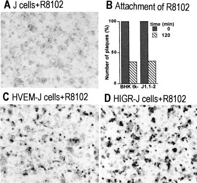
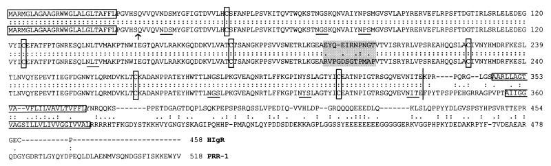
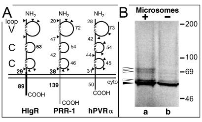
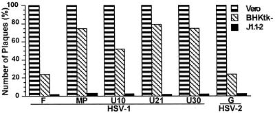
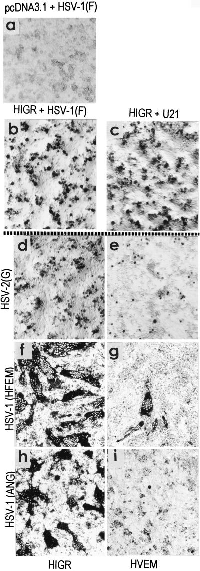
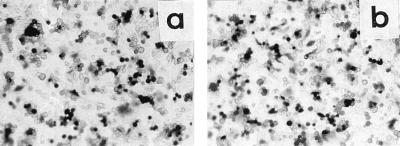
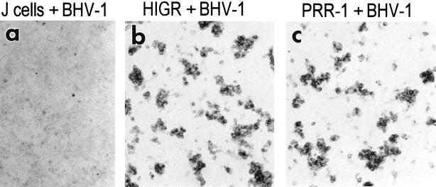
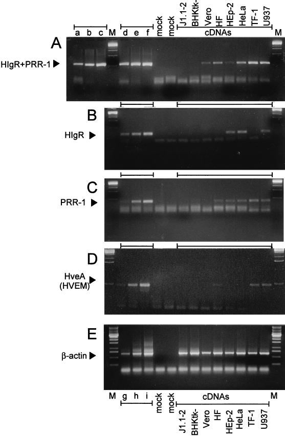
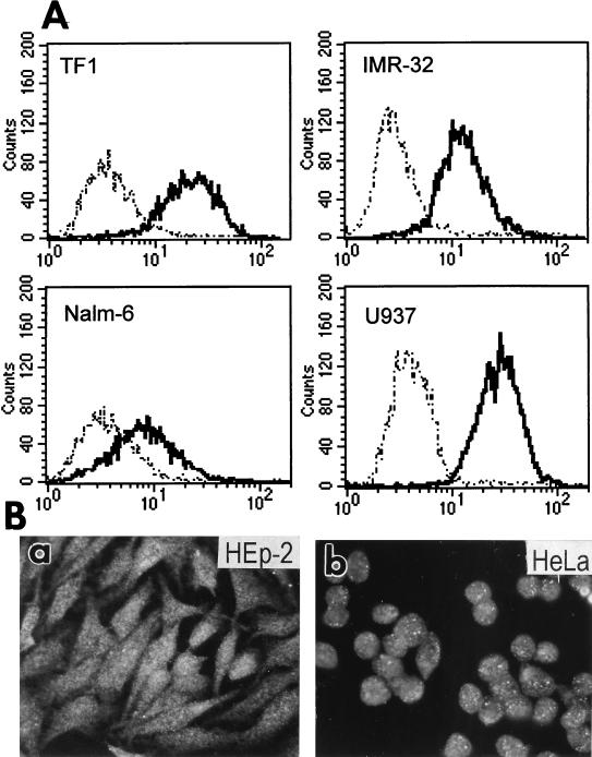
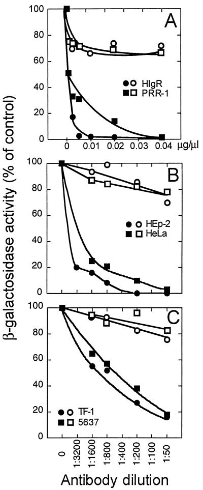
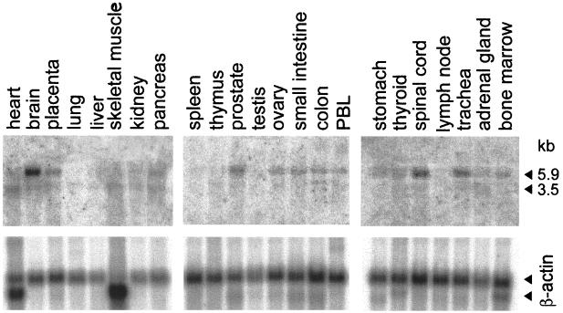
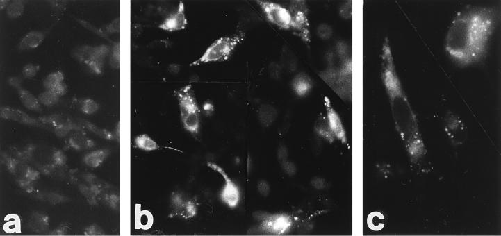
We report on the functional cloning of a hitherto unknown member of the immunoglobulin (Ig) superfamily selected for its ability to confer susceptibility to herpes simplex virus (HSV) infection on a highly resistant cell line (J1.1-2 cells), derived by exposure of BHKtk- cells to a recombinant HSV-1 expressing tumor necrosis factor alpha (TNF-alpha). The sequence of herpesvirus Ig-like receptor (HIgR) predicts a transmembrane protein with an ectodomain consisting of three cysteine-bracketed domains, one V-like and two C-like. HIgR shares its ectodomain with and appears to be an alternative splice variant of the previously described protein PRR-1 (poliovirus receptor-related protein). Both HIgR and PRR-1 conferred on J1.1-2 cells susceptibility to HSV-1, HSV-2, and bovine herpesvirus 1. The viral ligand of HIgR and PRR-1 is glycoprotein D, a constituent of the virion envelope long known to mediate viral entry into cells through interaction with cellular receptor molecules. Recently, PRR-1, renamed HveC (herpesvirus entry mediator C), and the related PRR-2, renamed HveB, were reported to mediate the entry of HSV-1, HSV-2, and bovine herpesvirus 1, and the homologous poliovirus receptor was reported to mediate the entry of pseudorabies virus (R. J. Geraghty, C. Krummenacher, G. H. Cohen, R. J. Eisenberg, and P. G. Spear, Science 280:1618-1620, 1998; M. S. Warner, R. J. Geraghty, W. M. Martinez, R. I. Montgomery, J. C. Whitbeck, R. Xu, R. J. Eisenberg, G. H. Cohen, and P. G. Spear, Virology 246:179-189, 1998). Here we further show that HIgR or PRR-1 proteins detected by using a monoclonal antibody to PRR-1 are widely distributed among human cell lines susceptible to HSV infection and commonly used for HSV studies. The monoclonal antibody neutralized virion infectivity in cells transfected with HIgR or PRR-1 cDNA, as well as in the human cell lines, indicating a direct interaction of virions with the receptor molecule, and preliminarily mapping this function to the ectodomain of HIgR and PRR-1. Northern blot analysis showed that HIgR or PRR-1 mRNAs were expressed in human tissues, with the highest expression being detected in nervous system samples. HIgR adds a novel member to the cluster of Ig superfamily members able to mediate the entry of alphaherpesviruses into cells. The wide distribution of HIgR or PRR-1 proteins among human cell lines susceptible to HSV infection, coupled with the neutralizing activity of the antibody in the same cells, provides direct demonstration of the actual use of this cluster of molecules as HSV-1 and HSV-2 entry receptors in human cell lines. The high level of expression in samples from nervous system makes the use of these proteins in human tissues very likely. This cluster of molecules may therefore be considered to constitute bona fide receptors for HSV-1 and HSV-2.


Figures

FIG. 1
FIG. 1
Characterization of J1.1-2 cells and identification of HIgR. (A) Micrograph of J1.1-2 (J) cells infected with the recombinant virus R8102 and stained for β-galactosidase to locate infected cells. No infected cell is visible. (B) Comparison of the extent of R8102 attachment to J1.1-2 and BHKtk− cells. Virus was allowed to attach to cells at 4°C; the virus present in the inoculum at zero time and that remaining unattached after 120 min were measured by a plaque assay in triplicate. (C and D) Micrographs of HveA(HVEM)-transfected (C) and HIgR-transfected (D) J1.1-2 cells infected with R8102 (5PFU/cell). X-Gal staining identified cells rendered susceptible by transfection.
FIG. 2
FIG. 2
Alignment of the HIgR and PRR-1 amino acid sequences by the ALIGN program, and relevant features of the proteins. The N-terminal signal sequences and transmembrane sequences are boxed, as are the six conserved cysteines involved in the formation of Ig domains. The arrow indicates the serine used for cleavage of the signal sequence. Potential N-glycosylation sites are underlined. The vertical line indicates the position of the splice site. The shaded box highlights the small divergent tract.
FIG. 3
FIG. 3
(A) Comparative schematic representation of the molecular structure of HIgR, PRR-1, and PVR. The V and C domains formed by cysteine bonds typical of this cluster of Ig superfamily members are shown, along with the number of residues in each region. The regions differing between HIgR and PRR-1, i.e., the transmembrane portion, the segment preceding it, and the cytoplasmic (cyto) tail, are in solid black for HIgR and shaded for PRR-1; the numbers of residues forming these latter regions are shown in bold type. Triangles indicate predicted N-glycosylation sites. (B) Electrophoretic mobility of HIgR synthesized by in vitro transcription-translation with 28.6 μCi of [35S]methionine (Radiochemical Center, Amersham, England) in the absence (lane b) or presence (lane a) of microsomes (Promega kit). Proteins were separated by denaturing electrophoresis in 8.5% polyacrylamide gels. The dried gel was analysed with a Bio-Rad PhosphoImager. The solid arrowhead points to unglycosylated precursor; the open arrowheads point to glycosylated species. The electrophoretic mobility of radioactive markers is shown on the right.
FIG. 4
FIG. 4
Relative plating efficiency of HSV-1 and HSV-2 strains in Vero, BHKtk−, and J1.1-2 cells. Plaques in Vero and BHKtk− cells were scored by Giemsa staining, and those in J1.1-2 cells were scored by immunostaining.
FIG. 5
FIG. 5
Effect of HIgR expression on the susceptibility of J1.1-2 cells to the indicated HSV-1 and HSV-2 strains. A gallery of representative micrographs of cells transfected with pcDNA3.1 vector alone (a), HIgR (b to d, f, and h), or HveA(HVEM) (e, g, and i) is shown. Infected cells were detected by immunostaining.
FIG. 6
FIG. 6
Transfection of J1.1-2 cells with PRR-1 cDNA confers susceptibility to R8102. Micrographs of J1.1-2 cells transfected with pLX1.12 (PRR-1) (a), or pCF18 (HIgR) (b), infected with R8102, and stained with X-Gal are shown.
FIG. 7
FIG. 7
Transfection of J1.1-2 cells with HIgR or PRR-1 confers susceptibility to BHV-1 infection. Micrographs of J1.1-2 cells transfected with pCF18 (HIgR) (b), pLX1.12 (PRR-1) (c), or pcDNA3.1 (a), infected with BHV-1, and immunostained with MAb 1240 to glycoprotein B are shown.
FIG. 8
FIG. 8
Agarose gel electrophoresis of RT-PCR-amplified sequences specific for HIgR plus PRR-1 (HIgR + PRR-1) (A), HIgR (B), PRR-1 (C), HveA(HVEM) (D), and β-actin (E). Sequences were amplified from cDNAs of the indicated cells (A to E) and from appropriate plasmids (A to D) or from cellular DNA (E). Each panel shows the intensity of amplified bands for increasing amounts of target sequences, as follows. (A) Lanes a to c and d to f contain 50, 500, and 5,000 copies of pLX1.12 (PRR-1) and pCF18 (HIgR), respectively. (B) Lanes d to f contain 5, 50, and 500 copies of pCF18. (C) Lanes d to f contain 50, 500, and 5,000 copies of pLX1.12. (D) Lanes d to f contain 50, 500, and 5,000 copies of pBEC10 for HveA. (E) Lanes g to i contain 0.1, 1, or 10 ng of DNA from human T lymphocytes. Lane M, molecular weight markers. Lanes mock, negative reaction controls lacking DNA but containing all other reagents. The sizes of the amplified products were 269, 119, 184, 233, and 674 bp for HIgR+PRR-1, HIgR, PRR-1, HveA, and β-actin, respectively. HF, MRC human fibroblasts.
FIG. 9
FIG. 9
Expression of HIgR and PRR-1 in human cell lines. (A) FACS analysis of TF-1, IMR-32, Nalm-6, and U937 cells. (B). Micrographs of HEp-2 (a) and HeLa (b) cells stained by indirect immunofluorescence with MAb R1.302 followed by biotinylated anti-mouse antibodies and Extravidin-TRITC.
FIG. 10
FIG. 10
Neutralization of R8102 infectivity by MAb R1.302 to PRR-1. (A) Stable transformants of J1.1-2 cells expressing HIgR or PRR-1 in 96-well plates were preincubated with the indicated dilutions (micrograms per microliters) of IgGs from MAb R1.302 to PRR-1 (solid symbols) or from purified mouse IgGs (open symbols) for 2 h at 4°C. R8102 was added to the antibody-containing medium and allowed to absorb for further 90 min at 4°C. (B) Infectivity neutralization in HEp-2 and HeLa cells with MAb R1.302 ascites fluid (solid symbols) or unrelated MAb (open symbols). (C) Infectivity neutralization in TF-1 and 5637 cells with MAb R1.302, ascites fluid (solid symbols) or unrelated MAb (open symbols).
FIG. 11
FIG. 11
Northern blot analysis of HIgR plus PRR-1 mRNA expression in human tissues. (A) Phosphoimages obtained with multiple-tissue Northern blot membranes hybridized with the BamHI fragment of pLX1.12 containing the entire PRR-1 cDNA. The probe did not distinguish between HIgR and PRR-1 mRNAs. Two bands, of 5.9 and 3.5 kbp, were detected. The highest level of expression occurred in brain and spinal cord tissues. (B) The same membranes hybridized with a β-actin probe.
FIG. 12
FIG. 12
Binding of gD to HIgR-expressing J1.1-2 cells. Fluorescence micrographs of J1.1-2 cells transfected with pCF18 (b and c) or pcDNA3.1 (a) are shown. Cells fixed with methanol were incubated with biotinylated recombinant gD-1(Δ290–299t) followed by Extravidin-TRITC.

References

    1. Arsenakis M, Campadelli Fiume G, Roizman B. Regulation of glycoprotein D synthesis: does alpha 4, the major regulatory protein of herpes simplex virus 1, regulate late genes both positively and negatively? J Virol. 1988;62:148–158. - PMC - PubMed
    1. Baines J D, Roizman B. The UL10 gene of herpes simplex virus 1 encodes a novel viral glycoprotein, gM, which is present in the virion and in the plasma membrane of infected cells. J Virol. 1993;67:1441–52. - PMC - PubMed
    1. Bonaldo M F, Lennon G, Soares M B. Normalization and subtraction: two approaches to facilitate gene discovery. Genome Res. 1996;6:791–806. - PubMed
    1. Brandimarti R, Huang T, Roizman B, Campadelli Fiume G. Mapping of herpes simplex virus 1 genes with mutations which overcome host restrictions to infection. Proc Natl Acad Sci USA. 1994;91:5406–5410. - PMC - PubMed
    1. Brunetti C R, Burke R L, Hoflack B, Ludwig T, Dingwell K S, Johnson D C. Role of mannose-6-phosphate receptors in herpes simplex virus entry into cells and cell-to-cell transmission. J Virol. 1995;69:3517–3528. - PMC - PubMed

Publication types

MeSH terms

Associated data

LinkOut - more resources