. 1998 Dec;72(12):10251-5.
doi: 10.1128/JVI.72.12.10251-10255.1998.

An endogenous inhibitor of human immunodeficiency virus in human lymphocytes is overcome by the viral Vif protein

Affiliations

An endogenous inhibitor of human immunodeficiency virus in human lymphocytes is overcome by the viral Vif protein

N Madani et al. J Virol. 1998 Dec.

Abstract

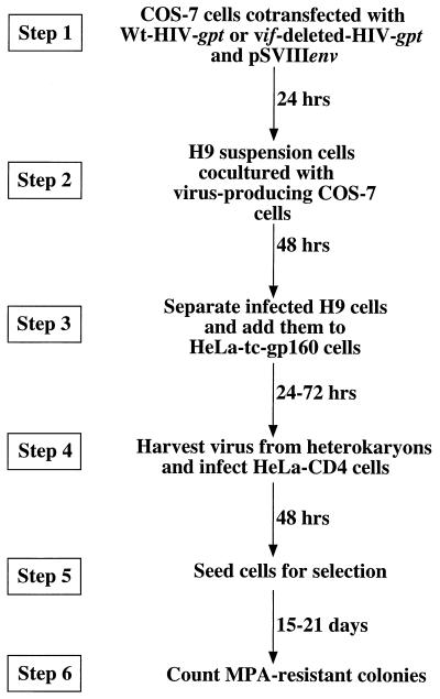
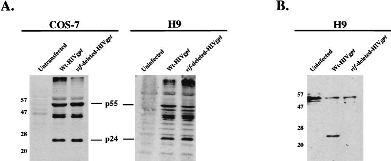
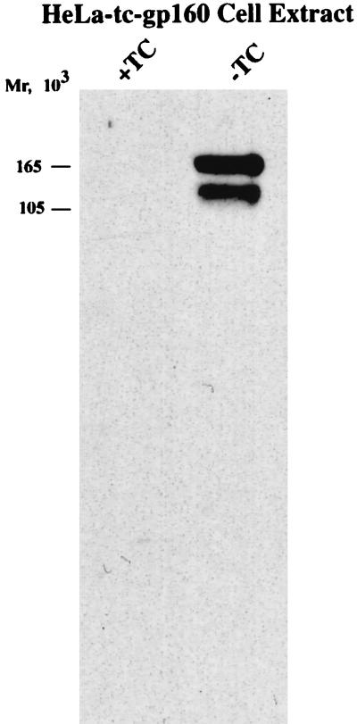
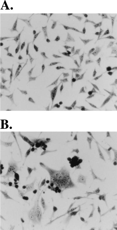
The vif gene of human immunodeficiency virus type 1 (HIV-1) encodes a basic Mr 23,000 protein that is necessary for production of infectious virions by nonpermissive cells (human lymphocytes and macrophages) but not by permissive cells such as HeLa-CD4. It had been proposed that permissive cells may contain an unidentified factor that functions like the viral Vif protein. To test this hypothesis, we produced pseudotyped wild-type and vif-deleted HIV gpt virions (which contain the HIV-1 genome with the bacterial mycophenolic acid resistance gene gpt in place of the viral env gene) in permissive cells, and we used them to generate nonpermissive H9 leukemic T cells that express these proviruses. We then fused these H9 cells with permissive HeLa cells that express the HIV-1 envelope glycoprotein gp120-gp41, and we asked whether the heterokaryons would release infectious HIV gpt virions. The results clearly showed that the vif-deleted virions released by the heterokaryons were noninfectious whereas the wild-type virions were highly infectious. This strongly suggests that nonpermissive cells, the natural targets of HIV-1, contain a potent endogenous inhibitor of HIV-1 replication that is overcome by Vif.


Figures

FIG. 1
FIG. 1
Schematic diagram of the vif complementation assay. In step 1, COS-7 cells at 80% confluency in a 100-mm culture dish were cotransfected with 5 μg of wild-type (Wt) pHIV-gpt or pHIVvif-deleted-gpt and 5 μg of pSVIIIenv. In step 2, suspensions of nonpermissive H9 leukemic T cells were added to the virus-producing COS-7 monolayers for 48 h. In step 3, the H9 cells were separated from the monolayer and cocultured with induced HeLa-tc-gp160 cells. This resulted in spontaneous cell fusion to produce heterokaryons, and virus was harvested from the culture media after 24, 48, and 72 h. In step 4, virus was filtered (0.45-μm-pore-size filters) and then used to infect HeLa-CD4 (H1-J clone) cells. In step 5, infected HeLa-CD4 cells were seeded in a 100-mm culture dish and selected with medium containing mycophenolic acid (MPA) (40 μg/ml). Mycophenolic acid-resistant colonies were fixed, stained, and counted after 15 to 21 days of selection (22).
FIG. 2
FIG. 2
Western immunoblot analyses of viral protein production in transfected COS-7 and infected H9 cells. H9 cells were cocultured with transfected virus-producing COS-7 cells as described in the legend to Fig. 1. At 72 h posttransfection of COS-7 cells (5 × 10⁶ cells per 100-mm culture dish) and 48 h postinfection of H9 cells (1 × 10⁶ cells), extracts of control and transfected or infected cells were obtained by washing the cells in phosphate-buffered saline (Life Technologies), followed by cell lysis in sodium dodecyl sulfate-polyacrylamide gel electrophoresis sample buffer (62.5 mM Tris-HCl [pH 6.8], 10% glycerol, 2% sodium dodecyl sulfate, 0.1% bromophenol blue, 10% 2-mercaptoethanol). The samples were then boiled, and equal amounts were loaded onto 10% polyacrylamide gels in the presence of 0.1% sodium dodecyl sulfate and subjected to electrophoresis. The proteins were then electrotransferred to nitrocellulose membranes and used for immunoblotting (21). Viral proteins were detected by incubating the membranes with HIV-IG antiserum (A) (obtained through the National Institutes of Health [NIH] AIDS Research and Reference Reagent Program, donated by Alfred Prince) or with HIV-1HXB2 Vif antiserum (B) (obtained through the NIH AIDS Research and Reference Reagent Program, donated by Dana Gabuzda) at a 1:1,000 dilution in 5% milk–0.1% Tween 20–Tris-buffered saline (Bio-Rad Laboratories, Hercules, Calif.), followed by protein A-conjugated horseradish peroxidase (HRP) at a 1:10,000 dilution (Bio-Rad). Antibody binding was then detected with a phototope-HRP Western blot detection kit (New England Biolabs). Mrs are indicated on the left, in thousands. Wt, wild type.
FIG. 3
FIG. 3
Western immunoblot analysis showing induced expression of gp160 upon removal of tetracycline (TC) from HeLa-tc-gp160 cells. HeLa-tc-gp160 cells were seeded in a 25-cm2 tissue culture flask and grown in the absence or presence of tetracycline (0.5 μg/ml; Sigma). Total cell extracts were collected 48 h later. Protein concentrations were measured by the Bio-Rad Bradford assay, and 20 μg of protein was analyzed by electrophoresis and immunoblotting, as described in the legend to Fig. 2. The blot was developed with a 1:1,000 dilution of sheep anti-gp120 antiserum (obtained through the NIH AIDS Research and Reference Reagent Program, donated by Michael Phelan), followed by protein G-conjugated horseradish peroxidase at a 1:5,000 dilution (Bio-Rad) and detection as described in the legend to Fig. 2.
FIG. 4
FIG. 4
Formation of syncytia by coculturing of HeLa-tc-gp160 and infected H9-42 (vif-deleted HIV gpt) cells. (A) HeLa-tc-gp160 cells grown in the presence of tetracycline do not synthesize gp160 or fuse with H9-42 cells. HeLa-tc-gp160 cells (3 × 10⁵) were seeded in a 25-cm2 tissue culture flask and grown for 2 days in medium containing 0.5 μg of tetracycline (Sigma) per ml before H9 cells (4 × 10⁶) were added. The cocultures were then grown for 48 h. Cells were observed under light microscopy after removal of the medium and unabsorbed cells, rinsing of adherent cells with phosphate-buffered saline (Life Technologies), fixing of cells with cold 100% methanol, and staining of cells with 0.1% toluidene blue in 30% ethanol. (B) H9 cells infected with vif-deleted HIV gpt were added to HeLa-tc-gp160 cells that were grown in the absence of tetracycline. Heterokaryons were allowed to form for 48 h. The cells were then fixed and stained as described for panel A. The small round darkly stained cells in both panels are H9 cells that adhered to the monolayers and were not removed by rinsing. Indistinguishable results were obtained with H9 cells that expressed wild-type HIV gpt (see text). Magnification, ×800.

References

    1. Ausubel F M, Brent R, Kingston R E, Moore D D, Seidman J G, Smith J A, Struhl K, editors. Current protocols in molecular biology. New York, N.Y: John Wiley & Sons, Inc.; 1994.
    1. Blanc D, Patience C, Schulz T F, Weiss R, Spire B. Transcomplementation of VIF− HIV-1 mutants in CEM cells suggests that VIF affects late steps of the viral life cycle. Virology. 1993;193:186–192. - PubMed
    1. Borman A M, Quillent C, Charneau P, Dauguet C, Clavel F. Human immunodeficiency virus type 1 Vif− mutant particles from restrictive cells: role of Vif in correct particle assembly and infectivity. J Virol. 1995;69:2058–2067. - PMC - PubMed
    1. Bouyac M, Rey F, Nascimbeni M, Courcoul M, Sire J, Blanc D, Clavel F, Vigne R, Spire B. Phenotypically Vif− human immunodeficiency virus type 1 is produced by chronically infected restrictive cells. J Virol. 1997;71:2473–2477. - PMC - PubMed
    1. Chen B K, Gandhi R T, Baltimore D. CD4 down-modulation during infection of human T cells with human immunodeficiency virus type 1 involves independent activities of vpu, env, and nef. J Virol. 1996;70:6044–6053. - PMC - PubMed

Publication types

MeSH terms

Substances