. 1998 Nov 10;95(23):13407-12.
doi: 10.1073/pnas.95.23.13407.

Propagating structure of Alzheimer's beta-amyloid(10-35) is parallel beta-sheet with residues in exact register

Affiliations

Propagating structure of Alzheimer's beta-amyloid(10-35) is parallel beta-sheet with residues in exact register

T L Benzinger et al. Proc Natl Acad Sci U S A. .

Abstract

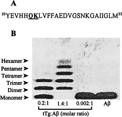
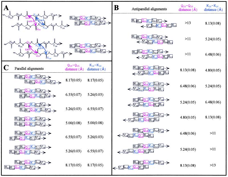
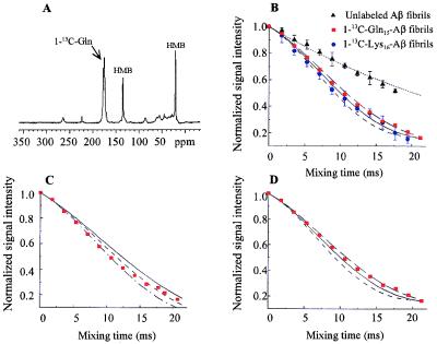
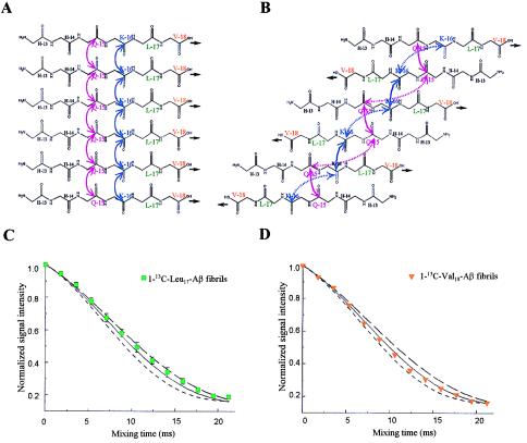
The pathognomonic plaques of Alzheimer's disease are composed primarily of the 39- to 43-aa beta-amyloid (Abeta) peptide. Crosslinking of Abeta peptides by tissue transglutaminase (tTg) indicates that Gln15 of one peptide is proximate to Lys16 of another in aggregated Abeta. Here we report how the fibril structure is resolved by mapping interstrand distances in this core region of the Abeta peptide chain with solid-state NMR. Isotopic substitution provides the source points for measuring distances in aggregated Abeta. Peptides containing a single carbonyl 13C label at Gln15, Lys16, Leu17, or Val18 were synthesized and evaluated by NMR dipolar recoupling methods for the measurement of interpeptide distances to a resolution of 0.2 A. Analysis of these data establish that this central core of Abeta consists of a parallel beta-sheet structure in which identical residues on adjacent chains are aligned directly, i. e., in register. Our data, in conjunction with existing structural data, establish that the Abeta fibril is a hydrogen-bonded, parallel beta-sheet defining the long axis of the Abeta fibril propagation.


Figures

Figure 1
Figure 1
Crosslinking by tTg suggests design of NMR experiments. (A) Amino acid sequence of Aβ(10–35). Peptides containing a 13C label at the carbonyl carbon of either Gln15 or Lys16 (underlined) were synthesized for use in solid-state NMR experiments. (B) Unlabeled Aβ(10–35) was crosslinked by tTg (69, 70) and was analyzed on Tris⋅Tricine SDS/PAGE (83). Reaction mixtures contained 1 mg/ml Aβ(10–35) peptide, 0.04–0.20 mg/ml tTg (Sigma), and 0.2–0.4 mM CaCl2 in 40 mM Tris (pH 8.6). After 4 h at 23°C, the experiments were stopped by addition of excess EDTA. Crosslinked peptide species ranged from dimeric to hexameric. The proportion of higher molecular weight species depended on the amount of tTg included; no higher order oligomers were observed. Specific crosslinking of Gln15 of one peptide to Lys16 of another, in a regular, ordered fashion, suggested placing 13C labels at these positions.
Figure 2
Figure 2
Modeling of β-strands indicating that orientation and alignment of Aβ can be determined by peptides containing single 1-13C labels in Gln15 or Lys16. (A) Top pair: the chemical and schematic views of a single alignment of two parallel β strands. The “pleat” of the β-pleated sheet is denoted by the offsets of the amino acids in the schematic view (right). For this single alignment, there are two possible offsets of the carbonyls, i.e., one where the carbonyls representing Gln15 (pink arrows) point out of the β-sheet (top pair) and one where the carbonyls of Gln15 point into the β-sheet (bottom pair). Interstrand carbonyl–carbonyl distances for parallel and antiparallel β-sheets were measured for >50 structures from the Brookhaven National Laboratory Protein Data Base, including isolated pairs of β-strands, sheets, and barrels. Two representative structures were chosen (46, 47). Shown in B are the measurements obtained for one antiparallel structure, in Å ± SE (n = 23–29), correlated with possible interstrand alignments of singly 1-13C labeled (Gln15 or Lys16) Aβ peptides. In C are the measurements obtained for one parallel structure, in Å ± SE (n = 34–38), correlated with possible interstrand alignments of singly 1-13C labeled (Gln15 or Lys16) Aβ peptides. The DRAWS experiment has a sensitivity limit of 6 Å and a precision of 0.1–0.2 Å; hence, it was clear at the outset of the experiment that if the Gln15 was indeed “near” the Lys16 of an adjacent peptide, combined distance data for the 1-13C-Gln15 and 1-13C-Lys16 peptides could be used to predict the form (parallel or antiparallel) and alignment of the peptides as they self-associate in the fibrils.
Figure 3
Figure 3
DRAWS data demonstrating a 5-Å, multiple-contact model for both 1-13C-Gln15-Aβ and 1-13C-Lys16-Aβ fibrils. (A) Conventional cross polarization/magic angle spinning experiment (DRAWS mixing time = 0) for 50 mg of 1-13C-Gln15-Aβ (δ = 171 ppm) mixed with 10 mg of unlabeled hexamethylbenzene (internal control). (B) For each peptide, the DRAWS experiment was performed by using a series of mixing times from 0 to 22 ms. At each mixing time, the carbonyl peak was integrated and normalized to the first data point (mixing time = 0) to allow comparison between samples. Data also were adjusted for natural abundance signal as described in Experimental Procedures. Values shown are the mean ± 1 SD for 50 mg of 1-13C-Gln15-Aβ lyophilized fibrils (red squares, n = 5), 15 mg of 1-13C-Lys16-Aβ (blue circles, n = 6), and 50 mg of unlabeled fibrils (black triangles, n = 5) compared with numerical simulations of no interaction (dotted line), a 5.2-Å interaction (long dashed line), a 5.0-Å interaction (solid line), or a 4.8-Å interaction (short dashed line). Error bars for experimental data are shown only when they exceed the symbol size. C shows the same data for 1-13C-Gln15-Aβ plotted against simulations for two 13C labels interacting in isolation (i.e., a single, two-spin contact) at 5.0 Å (solid line), 4.8 Å (short dashed line), or 4.6 Å (dot-dash line). D shows the 1-13C-Gln15-Aβ data plotted against simulations for three or more 13C labels interacting simultaneously (multiple spin, i.e., each 13C label has at least two contacts) at 5.2 Å (long dashed line), 5.0 Å (solid line), or 4.8 Å (short dashed line). At mixing times beyond 16 ms, the experimental data clearly fit a multiple-interaction model (D) better than an isolated-interaction model (C).
Figure 4
Figure 4
Further DRAWS experiments confirm parallel, directly aligned (“in register”) orientation of Aβ. Contacts (5 Å) for both Gln15 (pink) and Lys16 (blue) are consistent with either a parallel (A) or antiparallel (B) β-strand arrangement. In A, each 13C-labeled carbonyl has multiple interactions whereas in B, each 13C-labeled carbonyl has only paired interactions. The solid lines are contacts of ≈5 Å, and the dotted lines (B) are >8 Å, which would not be detectable in DRAWS experiments. Thus, the preferable fit of the data shown in Fig. 3D is consistent only with the parallel arrangement and not with the antiparallel arrangement. To validate this proposal, peptides containing 1-13C at Leu17 and Val18 were synthesized. A parallel arrangement (A) predicts 5-Å contacts for both peptides. An antiparallel arrangement (B) predicts that no measurable interaction at either Leu17 or Val18 would be found. (C) DRAWS experiments for 1-13C-Leu17-Aβ demonstrate a 5.1-Å (± 0.2) interaction at this position. Data shown are mean (± SD of mean) for two 50-mg samples, each run five times. (D) DRAWS experiments for 1-13C-Val18-Aβ demonstrate a 5.0-Å (± 0.2) interaction. Data shown are for one 50 mg-sample, run five times per data point. For both C and D, error bars are shown only when they exceed the size of the data point. Simulations shown are for multiple interactions (i.e., multiple spins) at 5.2-Å (long dashed line), 5.0-Å (solid line), or 4.8-Å (short dashed line).

References

    1. Glenner G G, Wong C W. Biochem Biophys Res Commun. 1984;122:1131–1135. - PubMed
    1. Masters C L, Simms G, Weinman N A, Multhaup G, McDonald B L, Beyreuther K. Proc Natl Acad Sci USA. 1985;82:4245–4249. - PMC - PubMed
    1. Kang J, Lemaire H G, Unterbeck A, Salbaum J M, Masters C L, Grzeschik K H, Multhaup G, Beyreuther K, Muller-Hill B. Nature (London) 1987;325:733–736. - PubMed
    1. Prelli F, Castaño E, Glenner G G, Frangione B. J Neurochem. 1988;51:648–651. - PubMed
    1. Levy E, Carman M D, Fernandez-Madrid I J, Power M D, Lieberburg I, van Duinen S G, Bots G T, Luyendijk W, Frangione B. Science. 1990;248:1124–1126. - PubMed

Publication types

Substances

LinkOut - more resources