. 1998 Nov 10;95(23):13513-8.
doi: 10.1073/pnas.95.23.13513.

The lipid phosphatase activity of PTEN is critical for its tumor supressor function

Affiliations

The lipid phosphatase activity of PTEN is critical for its tumor supressor function

M P Myers et al. Proc Natl Acad Sci U S A. .

Abstract

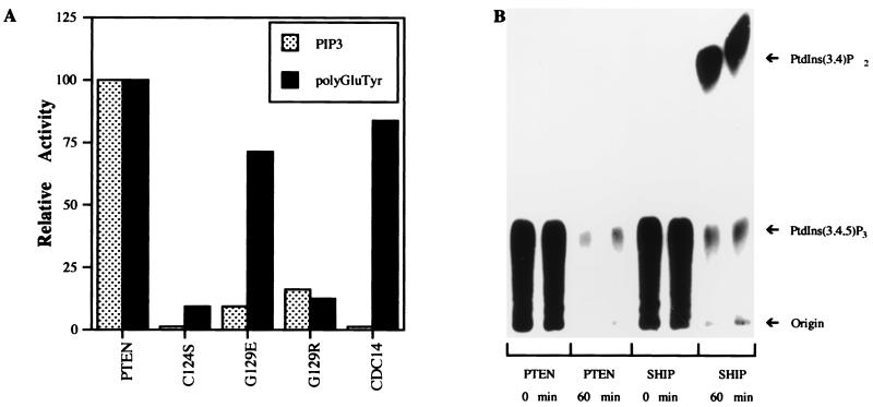
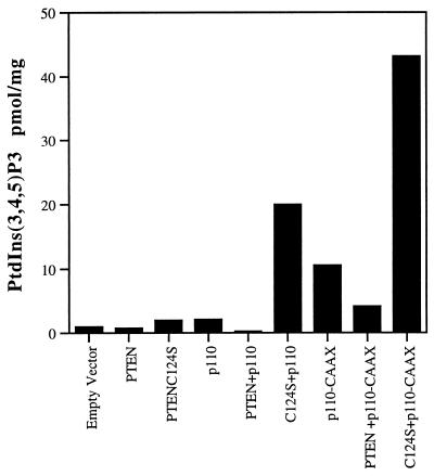
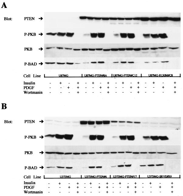
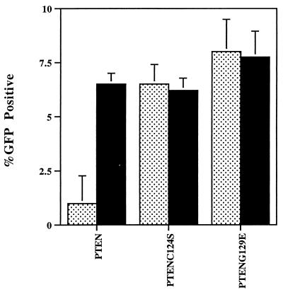
Since their discovery, protein tyrosine phosphatases have been speculated to play a role in tumor suppression because of their ability to antagonize the growth-promoting protein tyrosine kinases. Recently, a tumor suppressor from human chromosome 10q23, called PTEN or MMAC1, has been identified that shares homology with the protein tyrosine phosphatase family. Germ-line mutations in PTEN give rise to several related neoplastic disorders, including Cowden disease. A key step in understanding the function of PTEN as a tumor suppressor is to identify its physiological substrates. Here we report that a missense mutation in PTEN, PTEN-G129E, which is observed in two Cowden disease kindreds, specifically ablates the ability of PTEN to recognize inositol phospholipids as a substrate, suggesting that loss of the lipid phosphatase activity is responsible for the etiology of the disease. Furthermore, expression of wild-type or substrate-trapping forms of PTEN in HEK293 cells altered the levels of the phospholipid products of phosphatidylinositol 3-kinase and ectopic expression of the phosphatase in PTEN-deficient tumor cell lines resulted in the inhibition of protein kinase (PK) B/Akt and regulation of cell survival.


Figures

Figure 1
Figure 1
PTEN is a PtdIns 3-phosphatase. (A) Recombinant PTEN was incubated with radiolabeled polyGluTyr and PtdIns(3,4,5)P3, and the release of 32Pi was measured as described. (B) PtdIns(3,4,5)P3, labeled at the 3 position, was incubated with PTEN or SHIP for 0 or 60 min, and the reaction products were resolved on TLC plates and visualized by autoradiography.
Figure 2
Figure 2
Expression of PTEN-antagonized PI 3-kinase. HEK293 cells were transfected with PTEN or PTENC124S in combination with the p110 subunit of PI 3-kinase or an activated, membrane-bound PI-3 kinase (p110-CAAX). The resulting levels of PtdIns(3,4,5)P3 were determined as described and were normalized to total protein. Data are expressed as pmol of PtdIns(3,4,5)P3 per mg of protein. These data, from a single experiment, are representative of three experiments that yielded similar results.
Figure 3
Figure 3
Expression of PTEN in glioblastoma cell lines decreases the amount of activated PKB/AKT. PTEN expression was reconstituted in U87MG (A) or U373MB glioblastoma (B) cell lines by infection with recombinant retrovirus. Confluent dishes were either left untreated, stimulated with insulin (10 μg/ml for 10 min) or platelet-derived growth factor (50 ng/ml for 10 min), or pretreated with wortmannin (150 nM for 30 min), stimulated with both insulin (10 μg/ml) and platelet-derived growth factor (50 ng/ml) for 10 min, and then lysed. Equal amounts of protein were loaded for each cell line, and immunoblots were probed with antibodies to PTEN, PKB/Akt (PKB), phospho-PKB/Akt (P-PKB), or phosphorylated BAD (P-BAD) and visualized by using enhanced chemiluminescence reagents.
Figure 4
Figure 4
Expression of PTEN inhibits cell survival in LnCaP cells. PTEN, PTENC124S, or PTENG129E were cotransfected with a green fluorescent protein (GFP) expression vector into LnCaP cells. The cotransfections also included either an expression vector for consitutively active PKB/Akt (black bars) or a control empty vector (speckled bars). Transfected cells were identified as GFP positive cells or by immunofluorescence microscopy after staining with antibodies to the transfected PTEN. Transfection efficiency was assessed by determining the percentage of GFP/PTEN-positive cells (total number of cells was determined by counting nuclei stained with 4′,6-diamidino-2-phenylindole). Data are expressed as the mean transfection efficiency (± SD, n = 3).

References

    1. Steck P A, Perhouse M A, Jasser S A, Yung W K A, Lin H, Ligon A H, Lauren A L, Baumgard M L, Hattier T, Davis T, et al. Nat Genet. 1997;15:356–362. - PubMed
    1. Li J, Yen C, Liaw D, Podsypanina K, Bose S, Wang S, Puc J, Milliaresis C, Rodgers L, McCombie R, et al. Science. 1997;275:1943–1946. - PubMed
    1. Whang Y E, Wu X, Suzuki H, Reiter R E, Tran C, Vessella R L, Said J W, Isaacs W B, Sawyers C L. Proc Natl Acad Sci USA. 1998;95:5246–5250. - PMC - PubMed
    1. Rasheed B K, Stenzel T T, McLendon R E, Parsons R, Friedman A H, Friedman H S, Bigner D D, Bigner S H. Cancer Res. 1997;57:4187–4190. - PubMed
    1. Liaw D, Marsh D J, Li J, Dahia P L M, Wang S I, Zheng Z, Bose S, Call K M, Tsou H C, Peacocke M, et al. Nat Genet. 1997;16:64–67. - PubMed

Publication types

Substances