. 1998 Nov 10;95(23):13612-7.
doi: 10.1073/pnas.95.23.13612.

Impairing follicle-stimulating hormone (FSH) signaling in vivo: targeted disruption of the FSH receptor leads to aberrant gametogenesis and hormonal imbalance

Affiliations

Impairing follicle-stimulating hormone (FSH) signaling in vivo: targeted disruption of the FSH receptor leads to aberrant gametogenesis and hormonal imbalance

A Dierich et al. Proc Natl Acad Sci U S A. .

Abstract

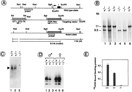
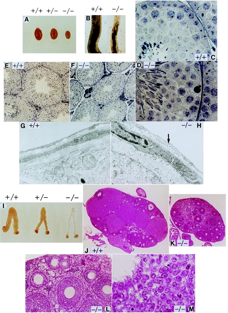
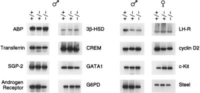
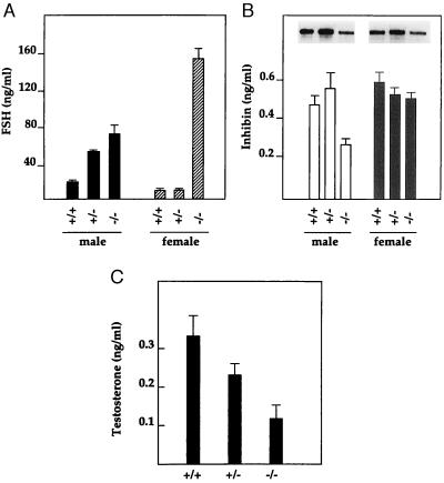
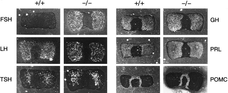
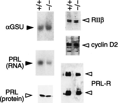
Pituitary gonadotropins follicle-stimulating hormone (FSH) and luteinizing hormone stimulate the gonads by regulating germ cell proliferation and differentiation. FSH receptors (FSH-Rs) are localized to testicular Sertoli cells and ovarian granulosa cells and are coupled to activation of the adenylyl cyclase and other signaling pathways. Activation of FSH-Rs is considered essential for folliculogenesis in the female and spermatogenesis in the male. We have generated mice lacking FSH-R by homologous recombination. FSH-R-deficient males are fertile but display small testes and partial spermatogenic failure. Thus, although FSH signaling is not essential for initiating spermatogenesis, it appears to be required for adequate viability and motility of the sperms. FSH-R-deficient females display thin uteri and small ovaries and are sterile because of a block in folliculogenesis before antral follicle formation. Although the expression of marker genes is only moderately altered in FSH-R -/- mice, drastic sex-specific changes are observed in the levels of various hormones. The anterior lobe of the pituitary gland in females is enlarged and reveals a larger number of FSH- and thyroid-stimulating hormone (TSH)-positive cells. The phenotype of FSH-R -/- mice is reminiscent of human hypergonadotropic ovarian dysgenesis and infertility.


Figures

Figure 1
Figure 1
Generation of FSH-R gene-deficient mice. (A) Strategy for generating the FSH-R null mutation. The EcoRV fragment containing exon 1 of the FSH-R gene was replaced by a PGK-Neo cassette. (B) DNA extracted from tail biopsies of wild-type (+/+), heterozygous (+/−), and homozygous (−/−) mice were digested with EcoRI; hybridization with probe 1 yielded 11-kb (wild type) and 9.5-kb (mutant) bands. (C) Northern analysis of poly(A)+ RNA extracted from mouse testis. (D) Reverse transcription–PCR from testis and ovary total RNA; primers used were selected within the sequence of exon 2 (sense primer: 5′-GTGCTCACCAAGCTTCGA-3′) and exon 7 (antisense primer: 5′-GAATCCCATTCTTATTCAGC-3′); all isoforms of the FSH-R are apparent. (E) FSH binding to crude membrane preparation obtained from testes of wild-type, heterozygous, and homozygous mice.
Figure 2
Figure 2
Analysis of testis and ovary from FSH-R mutant mice. (A) Testes comparison from 8-week-old wild-type, heterozygous, and homozygous littermates. (B) Transilluminated seminiferous tubules (stage VII–VIII of the seminiferous epithelium cycle) dissected from wild-type and homozygous mice; note the smaller tubule diameter in the mutant as compared with the wild type. Mutant mice conserve a normal spermatogenic wave. Histological sections at lower (E) and higher (C) magnification of seminiferous tubuli from a wild-type and mutant (D and F) mouse. (G and H) Electron micrographs of the basal membrane; note the altered organization of the collagen fibrils in mutant mouse tubules (arrow). (I) Reproductive tracts from 8-week-old littermates; ovaries and uteri from FSH-R mutant mice are smaller and thinner compared with the wild type; a reduction in size is also evident in the heterozygous. (J–M) Histological sections of the ovary from wild-type (J) and homozygous (K) mice. The decreased ovary size in the FSH-R −/− females is evident. There are no Graafian follicles nor corpora lutea. (L and M) Higher magnification of detailed structures in the FSH-R −/− ovary. Primary and secondary follicles appear normal (L), as well as the granulosa cells composition and organization (M). Magnifications: C and D ×370; E and F ×111; G and H ×18,000; J and K ×10; L ×96; M ×370.
Figure 3
Figure 3
Analysis of gene expression. Gonadal-specific gene expression evaluated by reverse transcription–PCR using 1-μg total RNA aliquots from 8-week-old wild-type, heterozygous, and homozygous mice. The identity of the amplified fragments was confirmed by Southern analysis hybridizing with internal probes.
Figure 4
Figure 4
Alteration in hormone levels. Serum from 8-week-old wild-type, heterozygous, and homozygous littermates was assayed for FSH, testosterone, and inhibin by RIA. Intracellular testosterone changes paralleled the variations in serum (not shown). (B) RNase protection assay of α-inhibin is shown.
Figure 5
Figure 5
Analysis of pituitary phenotype. Pituitary glands from 7-month-old wild-type and FSH-R mutant mice were fixed in OCT compound, and 10-μm cryostatic sections were prepared. The expression of the indicated gene was analyzed. Note the increased signal for FSH and TSH in mutant mice. GH, growth hormone; PRL, prolactin; POMC, proopiomelanocortin.
Figure 6
Figure 6
Expression analysis in the pituitary. Pituitary RNA (10 μg) was analyzed by Northern with GSUα and prolactin probes (23). Pituitary extracts were analyzed with a prolactin antibody (PRL); ovary extracts were analyzed with RIIβ, cyclin D2, and PRL-R antibodies.

References

    1. Ulloa-Aguirre A, Midgley A R, Beitins I Z, Padmanabhan V. Endocr Rev. 1995;16:765–787. - PubMed
    1. Sprengel R, Braun T, Nikolics K, Segaloff D, Seeburg P H. Mol Endocrinol. 1990;4:525–530. - PubMed
    1. Simoni M, Gromoll J, Nieschlag E. Endocr Rev. 1997;18:739–773. - PubMed
    1. Grasso P, Reichert L E. Endocrinology. 1990;128:949–956. - PubMed
    1. Sairam M R, Jiang L G, Yarney T A, Khan H. Biochem Biophys Res Comm. 1996;226:717–722. - PubMed

Publication types

Substances