. 1998 Nov 10;95(23):13618-23.
doi: 10.1073/pnas.95.23.13618.

Caspase-3 controls both cytoplasmic and nuclear events associated with Fas-mediated apoptosis in vivo

Affiliations

Caspase-3 controls both cytoplasmic and nuclear events associated with Fas-mediated apoptosis in vivo

T S Zheng et al. Proc Natl Acad Sci U S A. .

Abstract

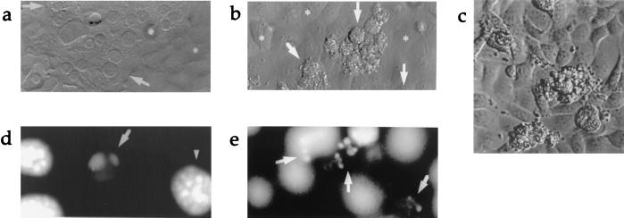
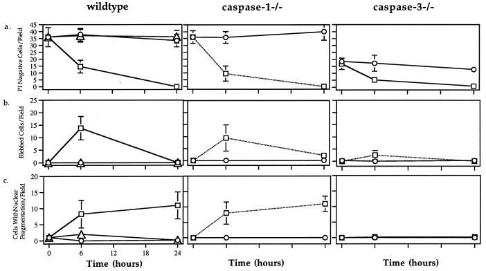
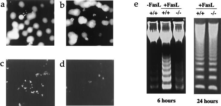
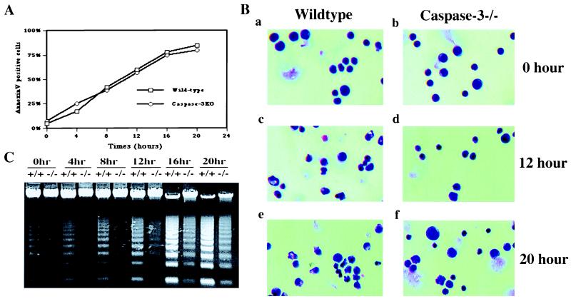
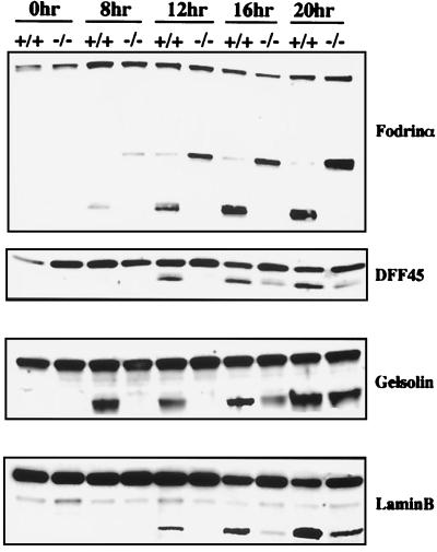
Both caspase-1- and caspase-3-like activities are required for Fas-mediated apoptosis. However, the role of caspase-1 and caspase-3 in mediating Fas-induced cell death is not clear. We assessed the contributions of these caspases to Fas signaling in hepatocyte cell death in vitro. Although wild-type, caspase-1(-/-), and caspase-3(-/-) hepatocytes were killed at a similar rate when cocultured with FasL expressing NIH 3T3 cells, caspase-3(-/-) hepatocytes displayed drastically different morphological changes as well as significantly delayed DNA fragmentation. For both wild-type and caspase-1(-/-) apoptotic hepatocytes, typical apoptotic features such as cytoplasmic blebbing and nuclear fragmentation were seen within 6 hr, but neither event was observed for caspase-3(-/-) hepatocytes. We extended these studies to thymocytes and found that apoptotic caspase-3(-/-) thymocytes exhibited similar "abnormal" morphological changes and delayed DNA fragmentation observed in hepatocytes. Furthermore, the cleavage of various caspase substrates implicated in mediating apoptotic events, including gelsolin, fodrin, laminB, and DFF45/ICAD, was delayed or absent. The altered cleavage of these key substrates is likely responsible for the aberrant apoptosis observed in both hepatocytes and thymocytes deficient in caspase-3.


Figures

Figure 1
Figure 1
(a) Light microscopic view (×600) of 24-hr coculture of wild-type hepatocytes and control fibroblasts. Arrows indicate normal hepatocytes. ∗ indicates fibroblast monolayer. (b) 24-hr coculture of wild-type hepatocytes with FasL-expressing fibroblasts (×600). Arrows point to dying hepatocytes. (c) Light microscopic view of blebbing hepatocytes with granular cytoplasm on the surface of monolayer of FasL-expressing fibroblasts (original magnification ×600, digitally enlarged). (d) DAPI (4′,6′-diamidino-2-phenylindole) staining illustrating hepatocyte nuclear condensation and (e) 24-hr coculture of wild-type hepatocytes with FasL-expressing fibroblasts (×320).
Figure 2
Figure 2
Time course of (a) cell death (PI negative cells), (b) blebbing, and (c) nuclear fragmentation of wild-type, caspase-1−/−, and caspase-3−/− hepatocytes after coculture with NIH 3T3 fibroblasts (○) or FasL-expressing NIH 3T3 fibroblasts with (▵) or without (□) caspase inhibitor z-VAD.fmk (10 μM).
Figure 3
Figure 3
DAPI (4′,6′-diamidino-2-phenylindole) staining illustrating typical small nuclear fragments in wild-type hepatocytes after coculture with FasL-expressing fibroblasts (a) and altered staining pattern in apoptotic caspase-3−/− hepatocytes (b). (c and d) Different TUNEL staining pattern in wild-type and caspase-3−/− hepatocytes 6 hr post-coculture. (e) Delayed DNA fragmentation in caspase-3−/− hepatocytes after coculture with FasL-expressing fibroblasts.
Figure 4
Figure 4
Kinetics of phosphatidylserine (PS) flip (A), nuclear morphology (B), and DNA fragmentation (C) in apoptotic wild-type and caspase-3−/− thymocytes. Thymocytes from either wild-type or caspase-3−/− mice were cultured in RPMI medium (10% fetal bovine serum) at 37°C in the presence of coated anti-Fas antibody Jo2. At each indicated time point, cells were harvested and subjected to morphological analysis, PS flipping, and DNA fragmentation assay as described in Materials and Methods.
Figure 5
Figure 5
Comparison of caspase substrate cleavage in wild-type and caspase-3−/− thymocytes during apoptosis. At each indicated time point after treatment (same as in Fig. 4), cells were lysed, and cleavage of various proteins were analyzed as described in Materials and Methods.

References

    1. Thompson C B. Science. 1995;267:1456–1462. - PubMed
    1. Steller H. Science. 1995;267:1445–1449. - PubMed
    1. Cohen G M. Biochem J. 1997;326:1–16. - PMC - PubMed
    1. Kuida K, Lippke J A, Ku G, Harding M W, Livingston D J, Su M S, Flavell R A. Science. 1995;267:2000–2003. - PubMed
    1. Li P, Allen H, Banerjee S, Franklin S, Herzog L, Johnston C, McDowell J, Paskind M, Rodman L, Salfeld J, et al. Cell. 1995;80:401–411. - PubMed

Publication types