. 1998 Nov 10;95(23):13698-702.
doi: 10.1073/pnas.95.23.13698.

Termination of signaling by protease-activated receptor-1 is linked to lysosomal sorting

Affiliations

Termination of signaling by protease-activated receptor-1 is linked to lysosomal sorting

J Trejo et al. Proc Natl Acad Sci U S A. .

Abstract

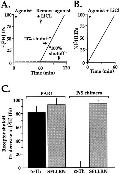
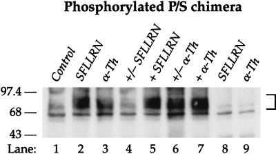
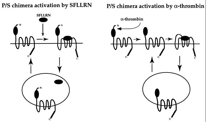
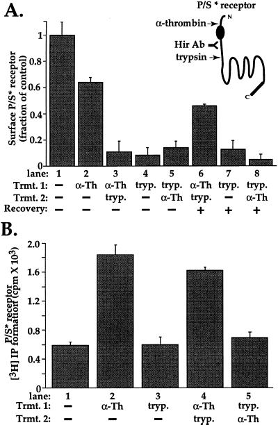
The irreversible proteolytic mechanism by which protease-activated receptor-1 (PAR1), the G protein-coupled receptor (GPCR) for thrombin, is activated raises the question of how it is shut off. Like classic GPCRs, activated PAR1 is rapidly phosphorylated and internalized, but unlike classic GPCRs, which recycle, internalized PAR1 is sorted to lysosomes. A chimeric PAR1 bearing the substance P receptor's cytoplasmic carboxyl tail sequestered and recycled like wild-type substance P receptor. In cells expressing this chimera, signaling in response to the PAR1-activating peptide SFLLRN ceased as expected upon removal of this agonist. Strikingly, however, when the chimera was activated proteolytically by thrombin, signaling persisted even after thrombin was removed. This persistent signaling was apparently due to "resignaling" by previously activated receptors that had internalized and recycled back to the cell surface. Thus the cytoplasmic carboxyl tail of PAR1 specifies an intracellular sorting pattern that is linked to its signaling properties. In striking contrast to most GPCRs, sorting of activated PAR1 to lysosomes rather than recycling is critical for terminating PAR1 signaling-a trafficking solution to a signaling problem.


Figures

Figure 1
Figure 1
Persistent signaling by P/S chimera after activation by thrombin but not SFLLRN. (A) Schematic of experimental design. Cells labeled with myo-[3H]inositol were incubated with agonist in the absence of LiCl; under these conditions, phosphoinositide hydrolysis is stimulated but IPs do not accumulate. After 60 min, agonist was removed and LiCl was added to allow accumulation of IPs during a subsequent incubation period as a measure of persistent signaling in the absence of agonist. Results were normalized to the amount of IPs accumulated when agonist and LiCl were added simultaneously (B) and expressed as percent shutoff (i.e., zero shutoff = no difference in IP accumulation under conditions A vs. B.) (C) Phosphoinositide hydrolysis was measured in PAR1-deficient mouse lung fibroblasts stably expressing similar amounts of surface wild-type PAR1 or the P/S chimera. myo-[3H]Inositol labeled cells were incubated with 100 μM SFLLRN or 10 nM α-Th for 60 min at 25°C in the absence of LiCl. Agonist was removed and cells were washed three times with DMEM containing 0.5 units/ml hirudin (α-Th inhibitor). Cells were then incubated with DMEM containing 20 mM LiCl and 0.5 units/ml hirudin at 25°C for an additional 60 min, at which time accumulated [3H]IPs were measured. The data are the mean % shutoff (see above) ± SD (n = 3). In PAR1-expressing cells, α-Th and SFLLRN caused an initial 3- and 3.8-fold increase in phosphoinositide hydrolysis, respectively; whereas cells expressing P/S chimera showed an initial 13- and 16-fold increase in PI hydrolysis to α-Th and SFLLRN, respectively. Similar results were obtained in three separate experiments. Note remarkable failure of α-Th-activated P/S chimera to shut off.
Figure 2
Figure 2
Proteolytically activated P/S chimera is continually phosphorylated even after thrombin is removed. Cells labeled with [32P]orthophosphate were treated with media alone (Control) or with 100 μM SFLLRN or 10 nM thrombin (α-Th) for either 3 (lanes 2 and 3) or 30 min (lanes 5 and 7, “+”). For reversible phosphorylation studies cells were incubated with agonists for 3 min, agonists were removed, cells were washed three times with DMEM/BSA and then incubated in media alone up to 30 min (lane 4 and 6, “+/−”). DMEM/BSA wash solution was supplemented with 0.5 units/ml hirudin for thrombin treated cells. Cell lysates were prepared and receptors were immunoprecipitated as described in Materials and Methods. Receptor immnoprecipitates were resolved by SDS/PAGE and analyzed by autoradiography. No phosphorylated receptor was detected in immunoprecipitates from agonist treated untransfected mouse lung fibroblasts (see lanes 8 and 9). Similar findings were observed in P/S chimera-expressing Rat1 fibroblasts.
Figure 3
Figure 3
Working model of reversible and irreversible activation of P/S chimera. When activated reversibly by agonist peptide SFLLRN, P/S chimera behaves like a classic GPCR. Activated P/S chimera is phosphorylated, internalized into an endosomal compartment where SFLLRN dissociates from receptor, then receptor recycles to the cell surface. When agonist is removed, receptors at the cell surface can enter their off state and receptors returning to the cell surface remain in their off state, thus signaling is effectively shutoff. When activated proteolytically by thrombin, P/S chimera follows the same trafficking route but because the tethered ligand remains present when receptor returns to the cell surface, P/S chimera becomes “reactivated” and signals again whether or not thrombin is present.
Figure 4
Figure 4
Receptor recycling and “reactivation” of signaling by proteolytically activated P/S chimera. (A) Recovery of proteolytically activated P/S* receptor at the cell surface was measured in stably transfected mouse lung fibroblasts by cell surface ELISA by using the antibody to the receptor’s hirudin-like domain (Hir Ab). Cells were incubated for 5 min at 25°C in the absence (lane 1) or presence (lane 2) of 10 nM α-Th, then fixed and the amount of receptor remaining on the cell surface was measured. Note the decrease in surface expression after α-Th, consistent with internalization of some receptors. For lanes 3–5 cells, cells were incubated with either 10 nM α-Th for 5 min at 25°C or with 100 nM trypsin (tryp.) for 20 min at 4°C. Proteases were then removed and cells were washed with DMEM containing either 0.5 units/ml hirudin or 2 μg/ml soybean trypsin inhibitor, then exposed a second time to either α-Th or trypsin as indicated. Cells were then fixed and the amount of receptor on the cell surface was determined. Note that trypsin effectively removed the hirudin-like domain epitope from the cell surface. For lanes 6–8, cells were treated as in lanes 3–5, but were incubated for an additional 60 min in the absence of proteases before the amount of P/S* receptor on the cell surface was measured. Note that significant recovery was seen only in cells previously exposed to α-Th (lane 6). The data shown are mean ± SD (n = 3) specific binding of hirudin-domain antibody to the cell surface; nonspecific antibody binding measured in untransfected cells was subtracted from total binding for each condition. Data are expressed as a fraction of specific binding measured in untreated cells (lane 1). Similar results were obtained in four separate experiments. (B) Signaling by P/S* receptor was measured in stably transfected lung fibroblasts labeled with myo-[3H]inositol. For lanes 2 and 3, cells were incubated in the presence or absence of 10 nM α-Th for 5 min at 25°C or 100 nM trypsin (tryp.) for 20 min at 4°C, proteases were then removed as above. For lanes 4 and 5, cells were exposed to α-Th and trypsin sequentially as indicated. For all lanes, LiCl was added after removal of the proteases, and accumulated [3H]IPs were measured after an additional 60 min incubation at 25°C. The data shown are the mean values ± SD (n = 3); basal [3H]IP formation was 590 cpm/well (lane 1). This experiment is representative of three independent experiments. Note that exposure to trypsin before but not after thrombin prevented persistent signaling.

References

    1. Yu S S, Lefkowitz R J, Hausdorff W P. J Biol Chem. 1993;268:337–341. - PubMed
    1. Krueger K M, Daaka Y, Pitcher J A, Lefkowitz R J. J Biol Chem. 1997;272:5–8. - PubMed
    1. Pippig S, Andexinger S, Lohse M J. Mol Pharmacol. 1995;47:666–676. - PubMed
    1. Freedman N J, Lefkowitz R J. Recent Prog Horm Res. 1996;51:319–351. ; discussion 352–353. - PubMed
    1. Hung D T, Vu T-K H, Wheaton V I, Ishii K, Coughlin S R. J Clin Invest. 1992;89:1350–1353. - PMC - PubMed

Publication types

MeSH terms

LinkOut - more resources