

Inactivating mutations in an SH2 domain-encoding gene in X-linked lymphoproliferative syndrome
- PMID: 9811875
- PMCID: PMC24894
- DOI: 10.1073/pnas.95.23.13765
Inactivating mutations in an SH2 domain-encoding gene in X-linked lymphoproliferative syndrome
Abstract
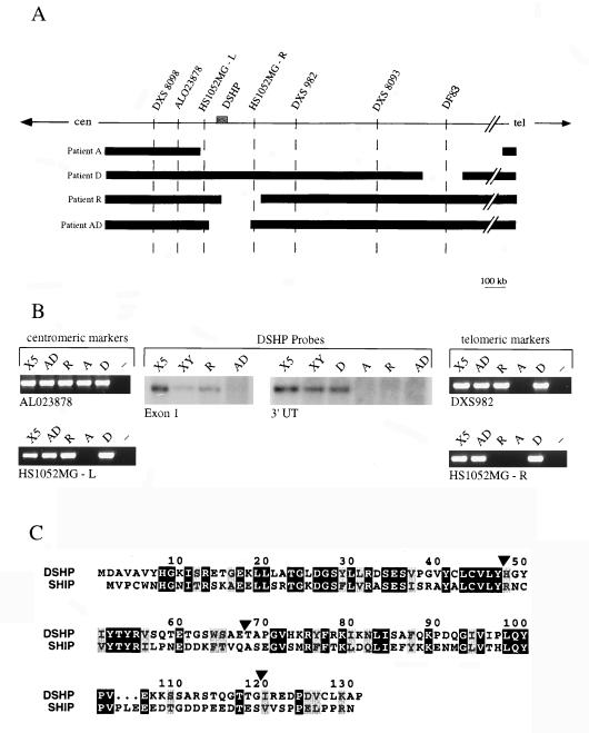
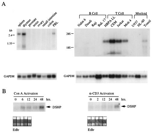
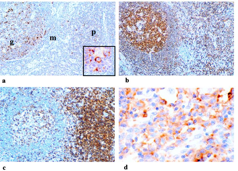
X-linked lymphoproliferative syndrome (XLP) is an inherited immunodeficiency characterized by increased susceptibility to Epstein-Barr virus (EBV). In affected males, primary EBV infection leads to the uncontrolled proliferation of virus-containing B cells and reactive cytotoxic T cells, often culminating in the development of high-grade lymphoma. The XLP gene has been mapped to chromosome band Xq25 through linkage analysis and the discovery of patients harboring large constitutional genomic deletions. We describe here the presence of small deletions and intragenic mutations that specifically disrupt a gene named DSHP in 6 of 10 unrelated patients with XLP. This gene encodes a predicted protein of 128 amino acids composing a single SH2 domain with extensive homology to the SH2 domain of SHIP, an inositol polyphosphate 5-phosphatase that functions as a negative regulator of lymphocyte activation. DSHP is expressed in transformed T cell lines and is induced following in vitro activation of peripheral blood T lymphocytes. Expression of DSHP is restricted in vivo to lymphoid tissues, and RNA in situ hybridization demonstrates DSHP expression in activated T and B cell regions of reactive lymph nodes and in both T and B cell neoplasms. These observations confirm the identity of DSHP as the gene responsible for XLP, and suggest a role in the regulation of lymphocyte activation and proliferation. Induction of DSHP may sustain the immune response by interfering with SHIP-mediated inhibition of lymphocyte activation, while its inactivation in XLP patients results in a selective immunodeficiency to EBV.
Figures
Comment in
-
DSHP: a "power bar" for sustained immune responses?Proc Natl Acad Sci U S A. 1998 Nov 10;95(23):13355-7. doi: 10.1073/pnas.95.23.13355. Proc Natl Acad Sci U S A. 1998. PMID: 9811804 Free PMC article. No abstract available.
References
-
- Straus S, Cohen J, Tosato G, Meier J. Ann Intern Med. 1993;118:45–58. - PubMed
-
- Callan M, Steven N, Krausa P, Wilson J, Moss P, Gillespie G, Bell J, Rickinson A, McMichael A. Nat Med. 1996;2:906–911. - PubMed
-
- Tosato G, Magrath I, Koski I, Dooley N, Blaese M. N Engl J Med. 1979;301:1133–1137. - PubMed
-
- Waele M D, Theilemans C, Camp B V. N Engl J Med. 1981;304:460–462. - PubMed
Publication types
MeSH terms
Substances
Grants and funding
LinkOut - more resources
Full Text Sources
Other Literature Sources
