. 1998 Nov 10;95(23):13829-34.
doi: 10.1073/pnas.95.23.13829.

Impaired liver regeneration in inducible nitric oxide synthasedeficient mice

Affiliations

Impaired liver regeneration in inducible nitric oxide synthasedeficient mice

R M Rai et al. Proc Natl Acad Sci U S A. .

Abstract

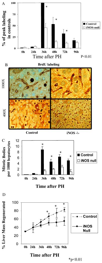
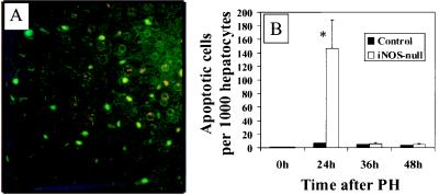
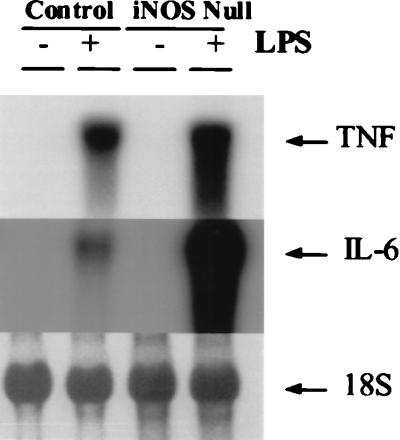
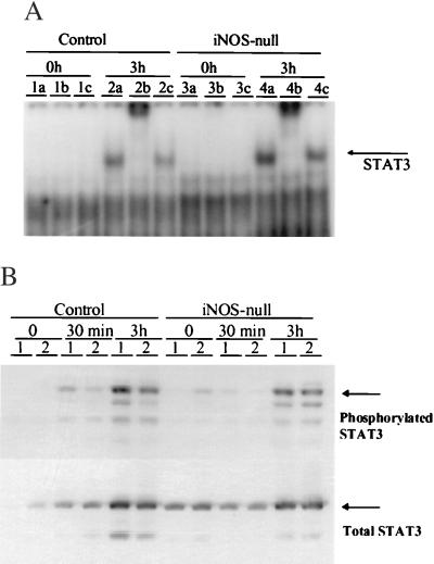
The mechanisms that permit adult tissues to regenerate when injured are not well understood. Initiation of liver regeneration requires the injury-related cytokines, tumor necrosis factor (TNF) alpha and interleukin (IL) 6, and involves the activation of cytokine-regulated transcription factors such as NF-kappabeta and STAT3. During regeneration, TNFalpha and IL-6 promote hepatocyte viability, as well as proliferation, because interventions that inhibit either cytokine not only block hepatocyte DNA synthesis, but also increase liver cell death. These observations suggest that the cytokines induce hepatoprotective factors in the regenerating liver. Given evidence that nitric oxide can prevent TNF-mediated activation of the pro-apoptotic protease caspase 3 and protect hepatocytes from cytokine-mediated death, cytokine-inducible nitric oxide synthase (iNOS) may be an important hepatoprotective factor in the regenerating liver. In support of this hypothesis we report that the hepatocyte proliferative response to partial liver resection is severely inhibited in transgenic mice with targeted disruption of the iNOS gene. Instead, partial hepatectomy is followed by increased caspase 3 activity, hepatocyte death, and liver failure, despite preserved induction of TNFalpha, IL-6, NF-kappabeta, and STAT3. These results suggest that during successful tissue regeneration, injury-related cytokines induce factors, such as iNOS and its product, NO, that protect surviving cells from cytokine-mediated death.


Figures

Figure 1
Figure 1
Liver regeneration is inhibited in iNOS-null mice. (A) The average number of BrdUrd-labeled hepatocytes was determined for each mouse by counting the number of positive hepatocytes in 10 different fields. Samples from 4–6 different mice/group were evaluated at each time point. Results shown are the mean ± SE of the averages from 4–6 mice/group per time point and are expressed as a percentage of the peak BrdUrd labeling in control mice. (B) Representative photomicrographs are shown from control and iNOS-deficient mice at two different magnifications [×400 (Lower) and ×1,000 (Upper)]. The dark-stained nuclei indicate BrdUrd-labeled hepatocytes. (C) Hepatocyte mitoses are expressed as the number of mitotic bodies/100 hepatocytes, and results are shown as the mean ± SE for each group. (D) Liver weights were used as another measure of liver regeneration. Results shown are the mean ± SE of data from 4–6 different mice/group per time point.
Figure 2
Figure 2
iNOS-deficient mice develop excessive lipid accumulation in hepatocytes after PH. Representative photomicrographs of hematoxylin/eosin-stained sections of livers from iNOS-deficient mice (A) and control mice (B) 48 hr after PH. (A.1 and B.1) Zone 3 (pericentral region) of the hepatic acinus. (A.2 and B.2) Mid-acinar region (zone 2). (A.3 and B.3) Zone 1 (periportal region) of the hepatic acinus. Note that mitotic figures (arrows) are easily identified in the periportal and mid-acinar regions of the livers of control mice but are not apparent when similar fields are inspected in iNOS-deficient livers. (Magnification = ×400.)
Figure 3
Figure 3
PH leads to foci of liver injury. Liver injury in iNOS-deficient mice may be manifest on hematoxylin/eosin-stained sections as (A) focal necrotic areas with hemorrhage and polymorphonuclear cell infiltration, (B) diffuse steatosis with inflammatory cell infiltration, or (C) distinct morphologic features of hepatocyte apoptosis with chromatin margination and/or condensation. (Magnification: A and C, ×400; B, ×1,000).
Figure 4
Figure 4
PH leads to hepatocyte apoptosis in iNOS-deficient mice. (A) Representative ×400 photomicrograph from TUNEL-stained liver sections from an iNOS-deficient mouse 24 hr after PH. Apoptotic cells are indicated by bright green fluorescence (FITC+). (B) Increased apoptotic activity in hepatocytes after PH in iNOS-deficient mice. A graphical representation of apoptotic activity in liver tissue sections obtained at 24, 36, and 48 hr from control and iNOS-deficient mice is shown. The percentage of hepatocytes with positive staining by TUNEL and classic features of chromatin margination and/or condensation and nuclear fragmentation are depicted on the x axis.
Figure 5
Figure 5
Macrophage production of TNFα and IL-6 is not decreased in iNOS-null mice. Autoradiographs of representative Northern blots for TNFα (Top) and IL-6 (Middle) are shown. (Bottom) The 18S RNA band on the same ethidium-bromide-stained blot.
Figure 6
Figure 6
Induction of STAT3 in iNOS-null mice at 3 hr after PH occurs normally, when compared with controls. (A) STAT3 DNA binding activity was determined by electrophoretic mobility-shift assay (EMSA) and supershift assays at various time points after PH. Maximal STAT3 binding activity occurred at 3 hr post-PH. This representative EMSA depicts STAT3 binding activity at baseline (time 0) and 3 hr after PH in controls and iNOS-null animals. Lanes 1 a-c contain time 0 extracts from control animals with added preimmune sera (a), total STAT3 antibody (b), or phosphorylated STAT3 antibody (c). Lanes 2 a-c contain extracts from controls at 3 hr post-PH in a similar order; lanes 3 a-c and 4 a-c contain extracts from iNOS-null animals at time 0 and 3 hr post-PH, respectively. (B) Total and phosphorylated STAT3 protein levels were determined by Western blot assay in control and iNOS-null mice at baseline (time 0), 30 min, and 3 hr after PH. Lanes 1 and 2 represent two individual animals used for the experiment at each time point.
Figure 7
Figure 7
PH induces proteolytic cleavage of a constitutively expressed 70-kDa hepatic protein in iNOS-null mice. To investigate hepatocyte apoptotic activity, in vivo cleavage of U1–70kD was assayed by immunoblotting with monospecific human sera recognizing U1–70kD. A signature 40-kDa peptide indicates apoptotic cleavage of the intact protein. Results shown are representative of those found in four different mice/group at 24 or 36 hr after PH. In this blot, each lane contains a sample from a different mouse at 36 hr after PH.

References

    1. Stocum D L. Science. 1997;276:15. - PubMed
    1. Tracey K J, Cerami A. Annu Rev Cell Biol. 1993;9:317–343. - PubMed
    1. Akerman P, Cote P, Yang S Q, McClain C, Nelson S, Bagby G J, Diehl A M. Am J Physiol. 1992;263:G579–G585. - PubMed
    1. Yamada Y, Kirillova I, Peschon J J, Fausto N. Proc Natl Acad Sci USA. 1997;94:1441–1446. - PMC - PubMed
    1. Romano M, Sironi M, Toniatti C, Polentarutti N, Fruscella P, Ghezzi P, Faggioni R, Luini W, van Hinsbergh V, Sozzani S, et al. Immunity. 1997;6:315–325. - PubMed

LinkOut - more resources