. 1998 Nov 10;95(23):13835-40.
doi: 10.1073/pnas.95.23.13835.

Dissociation between bone resorption and bone formation in osteopenic transgenic mice

Affiliations

Dissociation between bone resorption and bone formation in osteopenic transgenic mice

D A Corral et al. Proc Natl Acad Sci U S A. .

Abstract

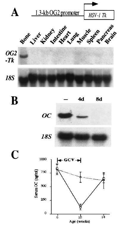
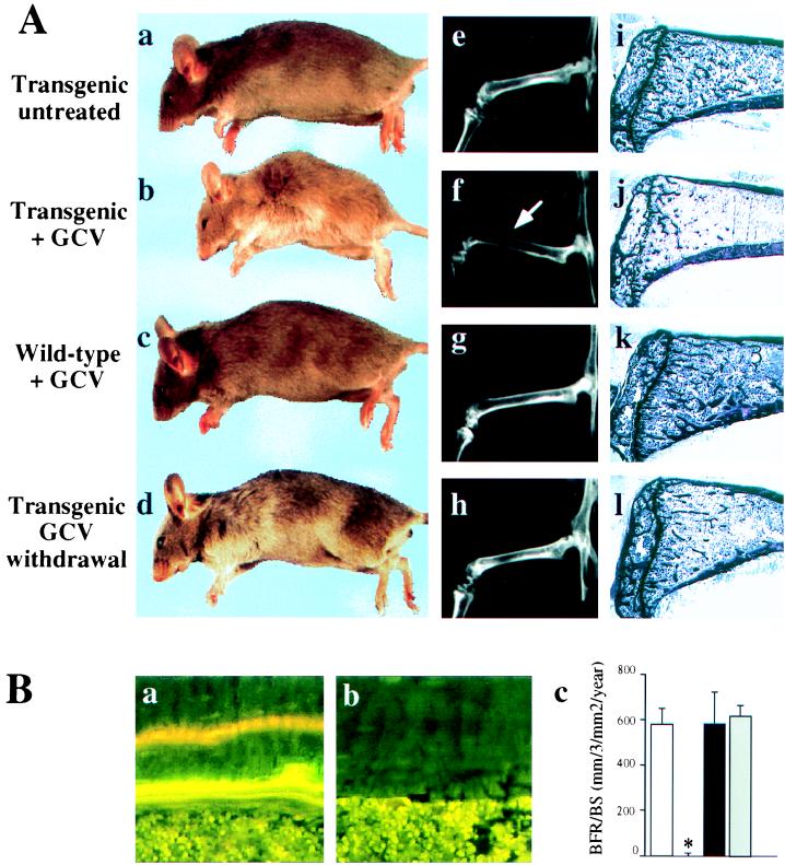
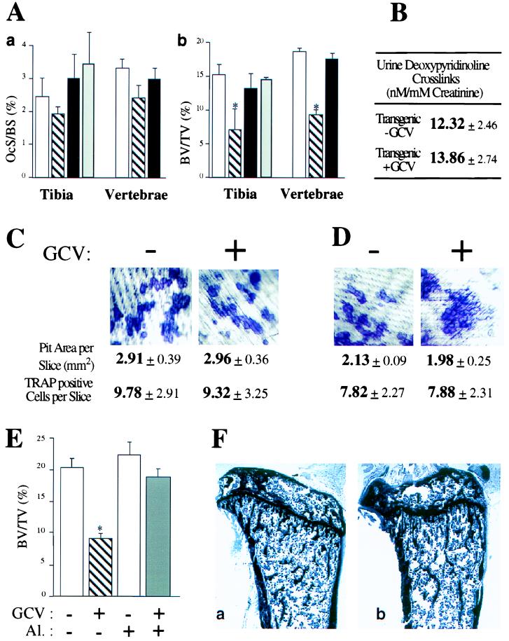
Bone mass is maintained constant in vertebrates through bone remodeling (BR). BR is characterized by osteoclastic resorption of preexisting bone followed by de novo bone formation by osteoblasts. This sequence of events and the fact that bone mass remains constant in physiological situation lead to the assumption that resorption and formation are regulated by each other during BR. Recent evidence shows that cells of the osteoblastic lineage are involved in osteoclast differentiation. However, the existence of a functional link between the two activities, formation and resorption, has never been shown in vivo. To define the role of bone formation in the control of bone resorption, we generated an inducible osteoblast ablation mouse model. These mice developed a reversible osteopenia. Functional analyses showed that in the absence of bone formation, bone resorption continued to occur normally, leading to an osteoporosis of controllable severity, whose appearance could be prevented by an antiresorptive agent. This study establishes that bone formation and/or bone mass do not control the extent of bone resorption in vivo.


Figures

Figure 1
Figure 1
Expression and activity of the OG2-Tk transgene. (A) Schematic representation of the transgene (Upper) and Northern blot analysis of its expression (Lower). (B) In vitro osteoblast ablation by GCV in calvarial cell culture of tg mice. Northern blot analysis shows osteocalcin (OC) expression in cells cultured without GCV for 4 days (−) and in the presence of 25 μM GCV for 4 days (4d) and 8 days (8d). Equivalent amounts of intact RNA were run in each lane as indicated by hybridization to an 18S RNA probe. (C) The graph shows in vivo osteoblast ablation after GCV treatment: measurement of circulating osteocalcin (OC) in wt animals (dotted line, n = 6) or tg animals (solid line, n = 6) before (6 weeks old), at the end of (10 weeks old), or 4 weeks after the GCV treatment period (14 weeks old).
Figure 2
Figure 2
Bone-formation parameters in GCV-treated animals. (A) Morphological, radiological, and histological comparison of 10-week-old tg mice untreated (a, e, and i) or treated daily for 4 weeks with GCV (b, f, and j), 10 week-old wt mice treated daily for 4 weeks with GCV (c, g, and k), or 14 week-old treated tg mice 4 weeks after GCV withdrawal (d, h, and l). Note the thinning of the cortices (arrow) and the lucent aspect of the bones on the x-rays (f) as well as the loss of the trabecular bone (j) in GCV-treated tg mice. These abnormalities were reversible after GCV withdrawal (compare f with h and j with l). (B) Dynamic histomorphometric analysis after tetracycline/calcein double labeling. Fluorescent micrographs of the two labeled mineralization fronts in representative section at the mid diaphysis of the tibia of wt (a) or tg mice (b) at the end of a 4-week GCV treatment period. (c) Measurement of the bone formation rate. Open bar, 10-week-old untreated tg mice; hatched bar, 10-week-old tg mice GCV-treated for 4 weeks; closed bar, 10-week-old wt mice GCV-treated for 4 weeks; gray bar, 14-week-old tg mice 4 weeks after GCV withdrawal. The asterisk indicates a statistically significant difference between wt and tg mice (P < 0.001; n = 4).
Figure 3
Figure 3
Bone resorption parameters in GCV-treated animals. (A) Measurement of osteoclast surface (a) and bone volumes (b) in tibia and vertebrae. Open bars, 10-week-old tg mice untreated; hatched bars, 10-week-old tg mice GCV-treated for 4 weeks; closed bars, 10-week-old wt mice GCV-treated for 4 weeks; gray bars, 14-week-old tg mice 4 weeks after GCV withdrawal. Asterisks indicate statistically significant differences following GCV treatment (P < 0.005; n = 4). (B) Urine deoxypyridinoline crosslinks levels in 10-week-old tg mice untreated or GCV-treated for 4 weeks. (C) Coculture of bone marrow cells (BMCs) and calvarial cells derived from tg mice treated in the absence (−) or presence (+) of GCV for 7 days. (D) Similar coculture assay of BMCs derived from 10-week-old tg mice treated for 4 weeks with GCV. (E) Measurement of bone volume after GCV and/or alendronate treatment of tg animals. Open bar, 10-week-old untreated mice; hatched bar, 10-week-old mice GCV-treated for 4 weeks; light gray bar, 10-week-old mice treated with alendronate (Al.) for 4 weeks; dark gray bar, 10-week-old mice treated with GCV and alendronate for 4 weeks. (F) Similar structural appearance of tibia of 10-week-old mouse untreated (a) or treated with GCV and alendronate for 4 weeks (b).
Figure 4
Figure 4
Induction of osteoporosis of variable severity. Tg animals aged 12 weeks were GCV-treated for 4–8 weeks. (A) Histological analysis of tibiae of 16-week-old mice untreated (a); 16-week-old mice GCV-treated for 4 weeks (b); 20-week-old mice GCV-treated for 8 weeks (c). Note the progressive loss of trabecular bone. (B) Static and dynamic histomorphometric analyses. Measurement of bone formation rate (a) and bone volume (b). Open bars, 16-week-old animal untreated; hatched bars, 16-week-old animals GCV-treated for 4 weeks; closed bars, 20-week-old animals GCV-treated for 8 weeks. For each parameter similar values were observed in 16-week-old and 20-week-old untreated animals. Asterisks indicate statistically significant differences between wt and tg mice (P < 0.01; n = 4).

Comment in

  • Bone homeostasis.
    Rodan GA. Rodan GA. Proc Natl Acad Sci U S A. 1998 Nov 10;95(23):13361-2. doi: 10.1073/pnas.95.23.13361. Proc Natl Acad Sci U S A. 1998. PMID: 9811806 Free PMC article. No abstract available.

References

    1. Frost H M. Calcif Tissue Res. 1969;3:211–237. - PubMed
    1. Hattner R, Epker B N, Frost H M. Nature (London) 1965;206:489–490. - PubMed
    1. Rodan G A, Martin T J. Calcif Tissue Int. 1981;33:349–351. - PubMed
    1. Rodan G A. In: Osteoporosis. Marcus R, Feldman D, Kelsey J, editors. San Diego: Academic; 1996. pp. 290–301.
    1. Lacey D L, Timms E, Tan H L, Kelley M J, Dunstan C R, Burgess T, Elliott R, Colombero A, Elliott G, Scully S, et al. Cell. 1998;93:165–176. - PubMed

Publication types

LinkOut - more resources