

Adeno-associated viral-mediated catalase expression suppresses optic neuritis in experimental allergic encephalomyelitis
- PMID: 9811889
- PMCID: PMC24923
- DOI: 10.1073/pnas.95.23.13847
Adeno-associated viral-mediated catalase expression suppresses optic neuritis in experimental allergic encephalomyelitis
Abstract
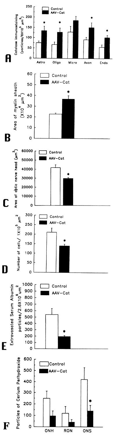
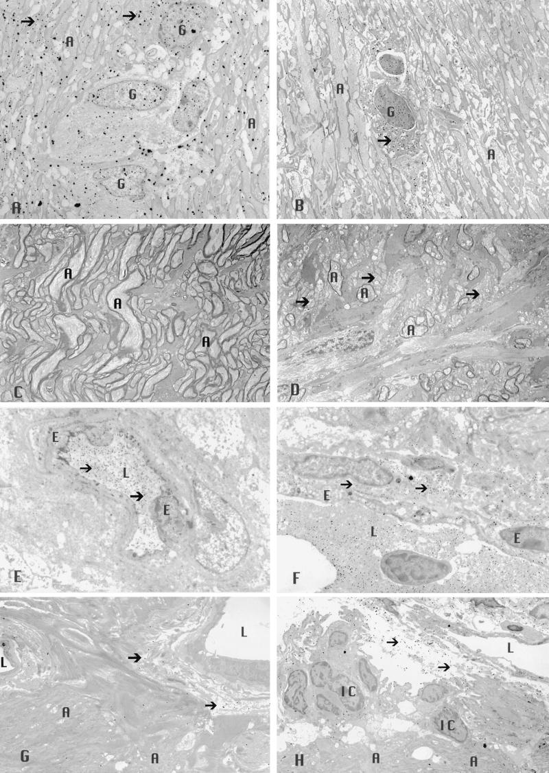
Suppression of oxidative injury by viral-mediated transfer of the human catalase gene was tested in the optic nerves of animals with experimental allergic encephalomyelitis (EAE). EAE is an inflammatory autoimmune disorder of primary central nervous system demyelination that has been frequently used as an animal model for the human disease multiple sclerosis (MS). The optic nerve is a frequent site of involvement common to both EAE and MS. Recombinant adeno-associated virus containing the human gene for catalase was injected over the right optic nerve heads of SJL/J mice that were simultaneously sensitized for EAE. After 1 month, cell-specific catalase activity, evaluated by quantitation of catalase immunogold, was increased approximately 2-fold each in endothelia, oligodendroglia, astrocytes, and axons of the optic nerve. Effects of catalase on the histologic lesions of EAE were measured by computerized analysis of the myelin sheath area (for demyelination), optic disc area (for optic nerve head swelling), extent of the cellular infiltrate, extravasated serum albumin labeled by immunogold (for blood-brain barrier disruption), and in vivo H2O2 reaction product. Relative to control, contralateral optic nerves injected with the recombinant virus without a therapeutic gene, catalase gene inoculation reduced demyelination by 38%, optic nerve head swelling by 29%, cellular infiltration by 34%, disruption of the blood-brain barrier by 64%, and in vivo levels of H2O2 by 61%. Because the efficacy of potential treatments for MS are usually initially tested in the EAE animal model, this study suggests that catalase gene delivery by using viral vectors may be a therapeutic strategy for suppression of MS.
Figures
References
-
- Raine C S. In: Handbook of Clinical Neurology. Vinken P J, Bruyn G W, Klawans H L, editors. Vol. 3. New York: Elsevier; 1985.
-
- Paterson P Y, Day E D. Am J Pathol. 1982;52:251–263.
-
- Rao N A. Invest Ophthalmol Visual Sci. 1981;20:159–172. - PubMed
-
- Beck R W, Cleary P A, Anderson M M, Jr, Keltner J L, Shults W T, Kaufman D I, Buckley E G, Corbett J J, Kupersmith M J, Miller N R, et al. N Engl J Med. 1992;326:581–588. - PubMed
-
- Rizzo J F, Lessell S. Neurology. 1988;38:185–190. - PubMed
Publication types
MeSH terms
Substances
Grants and funding
LinkOut - more resources
Full Text Sources
Other Literature Sources
