. 1998 Nov 10;95(23):13953-8.
doi: 10.1073/pnas.95.23.13953.

KATP channel inhibition by ATP requires distinct functional domains of the cytoplasmic C terminus of the pore-forming subunit

Affiliations

KATP channel inhibition by ATP requires distinct functional domains of the cytoplasmic C terminus of the pore-forming subunit

P Drain et al. Proc Natl Acad Sci U S A. .

Abstract

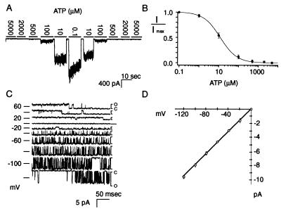
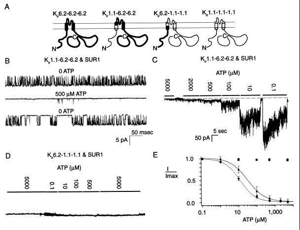
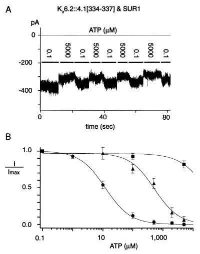
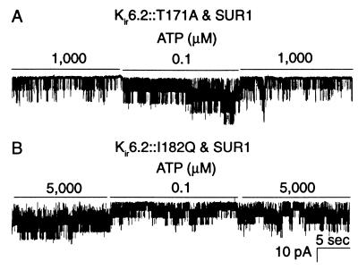
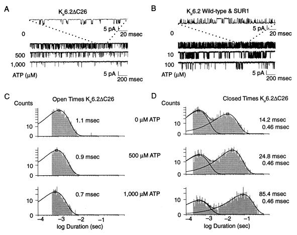
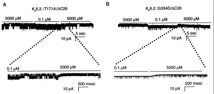
ATP-sensitive potassium ("KATP") channels are rapidly inhibited by intracellular ATP. This inhibition plays a crucial role in the coupling of electrical activity to energy metabolism in a variety of cells. The KATP channel is formed from four each of a sulfonylurea receptor (SUR) regulatory subunit and an inwardly rectifying potassium (Kir6.2) pore-forming subunit. We used systematic chimeric and point mutagenesis, combined with patch-clamp recording, to investigate the molecular basis of ATP-dependent inhibition gating of mouse pancreatic beta cell KATP channels expressed in Xenopus oocytes. We identified distinct functional domains of the presumed cytoplasmic C-terminal segment of the Kir6.2 subunit that play an important role in this inhibition. Our results suggest that one domain is associated with inhibitory ATP binding and another with gate closure.


Figures

Figure 1
Figure 1
Mouse SUR1 and mouse Kir6.2 coexpressed in Xenopus oocytes generate KATP channels. (A) Macroscopic currents from a representative inside-out patch in symmetrical 150 mM KCl at −80 mV. ATP at the indicated concentrations was superfused onto the cytoplasmic face of the patch. (B) Hill fit to data from eight oocytes. (C) Single channel currents at voltages from −120 to 60 mV in symmetrical 150 mM KCl and no ATP. (D) Linear fit to single-channel current amplitude as a function of voltage.
Figure 2
Figure 2
The N-terminal cytoplasmic segment of Kir6.2 is unlikely to be a major determinant of the ATP-dependent inhibition gating. Each construct was coexpressed with SUR1 and studied at −80 mV. (A) Parental and chimeric Kir constructs in which the N-terminal cytoplasmic segments are swapped. (B) Single channel records showing strong ATP sensitivity of channels formed from Kir1.1–6.2–6.2 and SUR1. (C) Corresponding macroscopic currents. (D) Macroscopic currents (≈730 pA) through channels formed from Kir6.2–1.1–1.1 show no ATP sensitivity. (E) Hill plots of ATP sensitivity of the chimeric constructs: •, wild-type Kir6.2 channels, Ki = 12.3 ± 3.5 μM and αH = 1.03 ± 0.10 (n = 8; taken from Fig. 1B); ▴, channels formed from Kir1.1–6.2–6.2, Ki = 30.0 ± 5.4 μM and αH = 1.0 ± 0.2 (n = 4); and ■, Kir6.2–1.1–1.1 (insensitive to ATP; n = 5).
Figure 3
Figure 3
Residues from the 334–337 region of Kir6.2 are required for wild-type ATP sensitivity. (A) Representative macroscopic current at −80 mV from a patch expressing channels formed from Kir6.2∷4.1[334–337] and SUR1 in response to alternating 0.1 and 5,000 μM ATP. The thin horizontal line indicates zero current. (B) ATP sensitivity of the 334–337 region chimeras: ▴, Kir6.2∷2.1[334–337] channels and ■, Kir6.2∷4.1[334–337] channels, compared with •, wild-type Kir6.2 channels, taken from Fig. 1B.
Figure 4
Figure 4
Kir6.2∷T171A and Kir6.2∷I182Q have low ATP sensitivity. (A) Representative single-channel currents at −80 mV from channels formed by coexpression of Kir6.2∷T171A and SUR1. The mutant channels exhibit moderate decrease in ATP sensitivity, exemplified by lowered but still substantial activity in the presence of 1,000 μM ATP. (B) Single channel currents at −80 mV from channels formed by Kir6.2∷I182Q and SUR1. The decrease in channel activity in response to low [ATP] is typical for this mutant. These channels are not detectably inhibited by ATP and appear to run down rapidly when [ATP] is below 10 μM.
Figure 5
Figure 5
Long-lived closed times of truncated Kir6.2ΔC26 largely fit into a single kinetic component and increase with [ATP]. (A) Truncated channel in the absence of SUR1. Top two traces in the absence of added ATP. Notice the very short bursts of openings and frequent long-lived closed times even in the absence of ATP. Bottom two traces at the [ATP] indicated. (B) Wild-type KATP channels. Notice the long bursts of openings and relatively rare long-lived closed times. (C) Open-time histograms of the truncated Kir6.2ΔC26 channel in the absence of SUR1 at the indicated [ATP], representative of four similar experiments. (D) Closed time histograms of the truncated Kir6.2ΔC26 channel in the absence of SUR1 at the indicated [ATP], representative of four similar experiments. The event duration histogram construction and fitting (12) results in a function of peaked exponential terms. The time constant of each term is specified by the x coordinate of the peak, indicated in the histograms by an open box. All currents were recorded at −80 mV.
Figure 6
Figure 6
On the ΔC26 background, T171A mutation restores bursting single-channel kinetics, whereas G334D mutation preserves spiking single- channel kinetics. (A) Kir6.2∷T171A/ΔC26 in the absence of SUR1, slow time scale, with a segment of the recording expanded on a fast time scale as indicated. On average, the 5,000 μM ATP inhibits one of two channels. Notice that the T171A mutation reverts the spiking single-channel gating kinetics of the ΔC26 parent channel to the long bursts of rapid successive openings similar to the wild-type KATP channel (see Fig. 5B). (B) Kir6.2∷G334D/ΔC26 in the absence of SUR1, slow time scale, with a segment of the recording expanded on a fast time scale as indicated. Notice that the channels retain the spiking single-channel gating kinetics of the ΔC26 parent channel. (See Fig. 5A). All currents were recorded at −80 mV.

References

    1. Inagaki N, Gonoi T, Clement J P, IV, Namba N, Inazawa J, Gonzalez G, Aguilar-Bryan L, Seino S, Bryan J. Science. 1995;270:1166–1170. - PubMed
    1. Clement J P, IV, Kunjilwar K, Gonzales G, Schwanstecher M, Panten U, Aguilar-Bryan L, Bryan J. Neuron. 1997;18:827–838. - PubMed
    1. Inagaki N, Gonoi T, Seino S. FEBS Lett. 1997;409:232–236. - PubMed
    1. Shyng S-L, Nichols C G. J Gen Physiol. 1997;110:655–664. - PMC - PubMed
    1. Aguilar-Bryan L, Nichols C G, Wechsler S W, Clement J P T, IV, Boyd A E, Gonzalez G, Herrera-Sosa H, Nguy K, Bryan J, Nelson D A. Science. 1995;268:423–426. - PubMed

Publication types

LinkOut - more resources