. 1998 Nov 10;95(23):13976-81.
doi: 10.1073/pnas.95.23.13976.

Eukaryotic phytochromes: light-regulated serine/threonine protein kinases with histidine kinase ancestry

Affiliations

Eukaryotic phytochromes: light-regulated serine/threonine protein kinases with histidine kinase ancestry

K C Yeh et al. Proc Natl Acad Sci U S A. .

Abstract

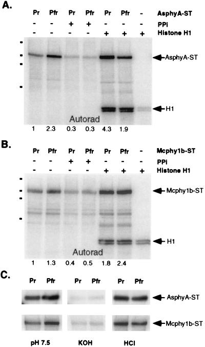
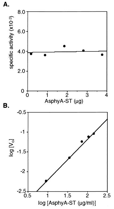
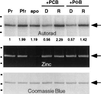
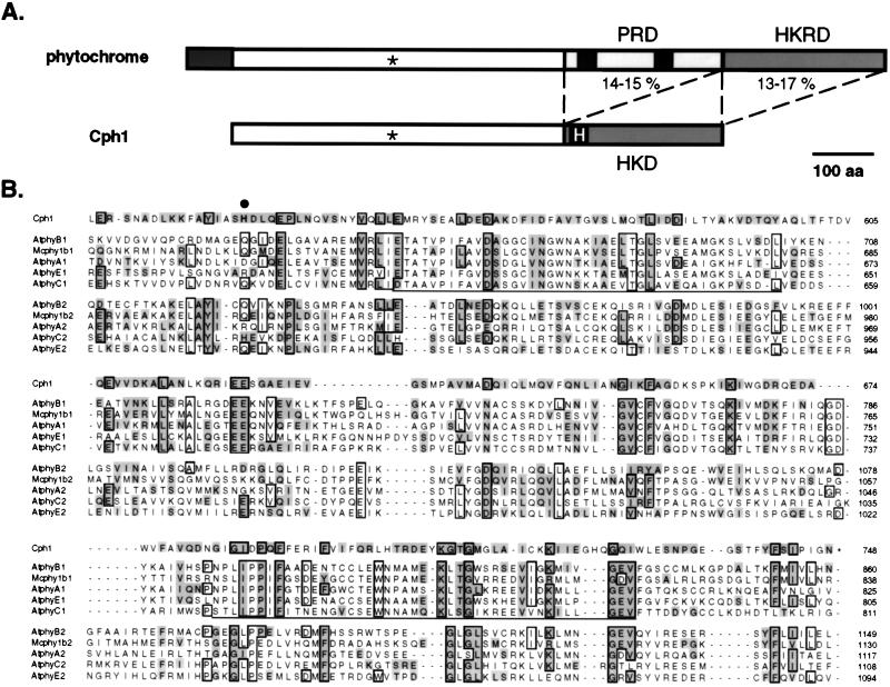
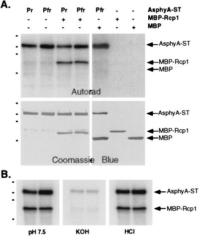
The discovery of cyanobacterial phytochrome histidine kinases, together with the evidence that phytochromes from higher plants display protein kinase activity, bind ATP analogs, and possess C-terminal domains similar to bacterial histidine kinases, has fueled the controversial hypothesis that the eukaryotic phytochrome family of photoreceptors are light-regulated enzymes. Here we demonstrate that purified recombinant phytochromes from a higher plant and a green alga exhibit serine/threonine kinase activity similar to that of phytochrome isolated from dark grown seedlings. Phosphorylation of recombinant oat phytochrome is a light- and chromophore-regulated intramolecular process. Based on comparative protein sequence alignments and biochemical cross-talk experiments with the response regulator substrate of the cyanobacterial phytochrome Cph1, we propose that eukaryotic phytochromes are histidine kinase paralogs with serine/threonine specificity whose enzymatic activity diverged from that of a prokaryotic ancestor after duplication of the transmitter module.


Figures

Figure 1
Figure 1
Recombinant oat and algal phytochromes possess protein kinase activity. (A) Autoradiograph of Pr and Pfr forms of PCB adducts of recombinant oat phytochrome A (AsphyA-ST; 0.5 μg) after incubation with radiolabeled ATP under standard kinase conditions in the presence (+) or absence (−) of 2 μg histone H1 and/or 2 mM pyrophosphate (PPi) was obtained as described in Materials and Methods. The relative amount of phosphorylation indicated below the autoradiographs was obtained by using imagequant software (Molecular Dynamics). Dots on the left side of the gel represent molecular mass standards with 197 kDa, 117 kDa, 89 kDa, and 52 kDa (top to bottom). (B) An autoradiograph of algal phytochrome (Mcphy1b-ST; 1 μg) assayed identically as AsphyA-ST (above). (C) Hydrolytic stability of autophosphorylated phytochromes examined by treatment of poly(vinylidene difluoride) membranes with neutral (50 mM Tris⋅HCl, pH 7.5), basic (3 M KOH), or acidic (1 M HCl) solutions for 2.5 hr at room temperature with gentle shaking.
Figure 2
Figure 2
ATP-dependent phosphorylation of the PCB adduct of recombinant oat phytochrome is an intramolecular process. (A) Specific activity of AsphyA-ST autophosphorylation (dpm/μg AsphyA-ST; Pfr form) plotted versus μg AsphyA-ST analyzed. (B) A van’t Hoff plot of the same data obtained by plotting the logarithm of the initial rate (Vo) of AsphyA-ST phosphorylation, determined by converting the phosphorylation level (dpm) to pmol incorporation per minute, versus the logarithm of AsphyA-ST concentration (μg/ml). The slope of this line was 1.02 (r2 = 0.994).
Figure 3
Figure 3
Phytochrome autophosphorylation is both chromophore and light regulated. Autoradiograph (Top), zinc blot (Middle), and Coomassie blue-stained blot (Bottom) of an AsphyA-ST apoprotein sample divided into three fractions. One fraction was treated with dimethyl sulfoxide only (apo), and the other two were treated with 8 μM PCB and 8 μM phytochromobilin (PΦB) and incubated 30 min in complete darkness, and assayed for kinase activity as described in Materials and Methods. Apophytochrome (apo), newly assembled bilin-adducts (D), and red-light irradiated bilin adducts (R) are shown (0.5 μg). Preassembled Pr and Pfr forms of AsphyA-ST (PCB adduct; 0.5 μg) were included in the left two lanes for comparison. Relative phosphorylation and molecular mass markers were determined as in Fig. 1.
Figure 4
Figure 4
Duplication of a transmitter module of a prokaryotic phytochrome ancestor: A model for molecular evolution of eukaryotic phytochromes. (A) Structural comparison of eukaryotic phytochromes and Cph1. The conserved cysteine chromophore binding site is marked (∗), and the conserved histidine on the HKD of Cph1 is highlighted. The percent amino acid equivalence between the HKD of Cph1 and both PRD and HKRD of eukaryotic phytochromes is indicated (see Table 1 for details). (B) Multiple sequence alignment of the HKD of Cph1 with the PRD (sequences ending with 1) and HKRD (sequences ending with 2) regions of representative eukaryotic phytochromes including phytochromes from Arabidopsis thaliana (i.e., AtphyA, AtphyB, AtphyC, and AtphyE) and phytochrome from the green alga M. caldariorum. (Mcphy1b). Residues that are identical in six or more sequences are boxed, whereas residues equivalent to those in Cph1 (where I = V, L = M, R = K, S = T = A and D = E = N = Q) are highlighted. The conserved histidine phosphorylation site (H538) of Cph1 is marked with a large dot. The two PAS motifs in eukaryotic phytochromes are underlined. GenBank accession numbers for phytochrome sequences are AtphyA, L21154; AtphyB, X17342; AtphyC, Z32538; AtphyE, X76610; and Mcphy1b, U31284.
Figure 5
Figure 5
MBP-Rcp1 is a substrate for AsphyA-ST kinase activity. (A) Autoradiograph and Coomassie blue-stained blot of Pr and Pfr forms of AsphyA-ST (PCB adduct; 1 μg) incubated alone or with MBP-Rcp1 (1 μg) under standard kinase assay conditions (see Materials and Methods). As controls, a mixture of purified MBP (1.5 μg) and the Pfr form of AsphyA-ST (PCB adduct; 1 μg), purified MBP-RCP1 (1 μg) or purified MBP (1.5 μg) incubated under standard kinase assay conditions (see Materials and Methods) are shown in the right three lanes. (B) Acid and base hydrolytic stability of the mixture of AsphyA-ST (PCB adduct, 1 μg) and MBP-Rcp1 (1 μg) was determined as described in Fig. 1.

Comment in

Similar articles

Cited by

References

    1. Sage L C. Pigment of the Imagination: A History of Phytochrome Research. San Diego: Academic; 1992.
    1. Smith H. Annu Rev Plant Physiol Plant Mol Biol. 1995;46:289–315.
    1. Pratt L H. Photochem Photobiol. 1995;61:10–21. - PubMed
    1. Furuya M, Schafer E. Trends Plant Sci. 1996;1:301–307.
    1. Quail P H. Plant Cell Environ. 1997;20:657–665.

Publication types

MeSH terms

LinkOut - more resources