. 1998 Nov 13;282(5392):1327-32.
doi: 10.1126/science.282.5392.1327.

A structural explanation for the recognition of tyrosine-based endocytotic signals

Affiliations

A structural explanation for the recognition of tyrosine-based endocytotic signals

D J Owen et al. Science. .

Abstract

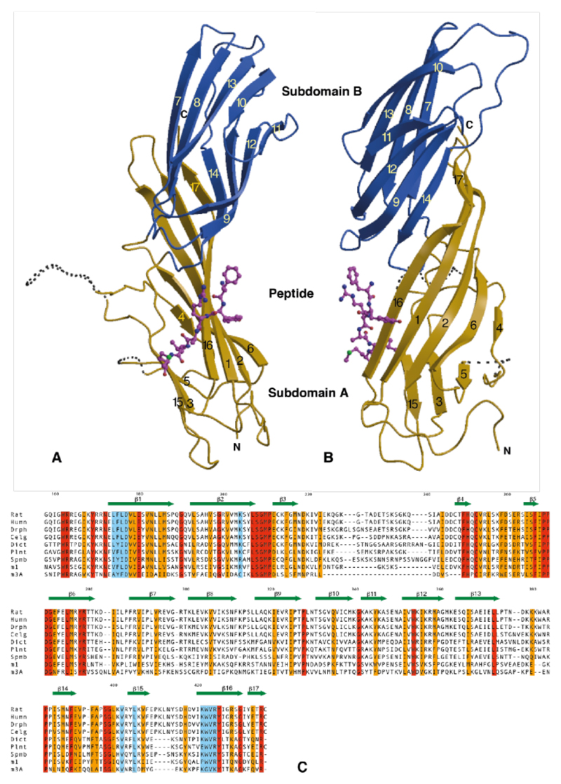
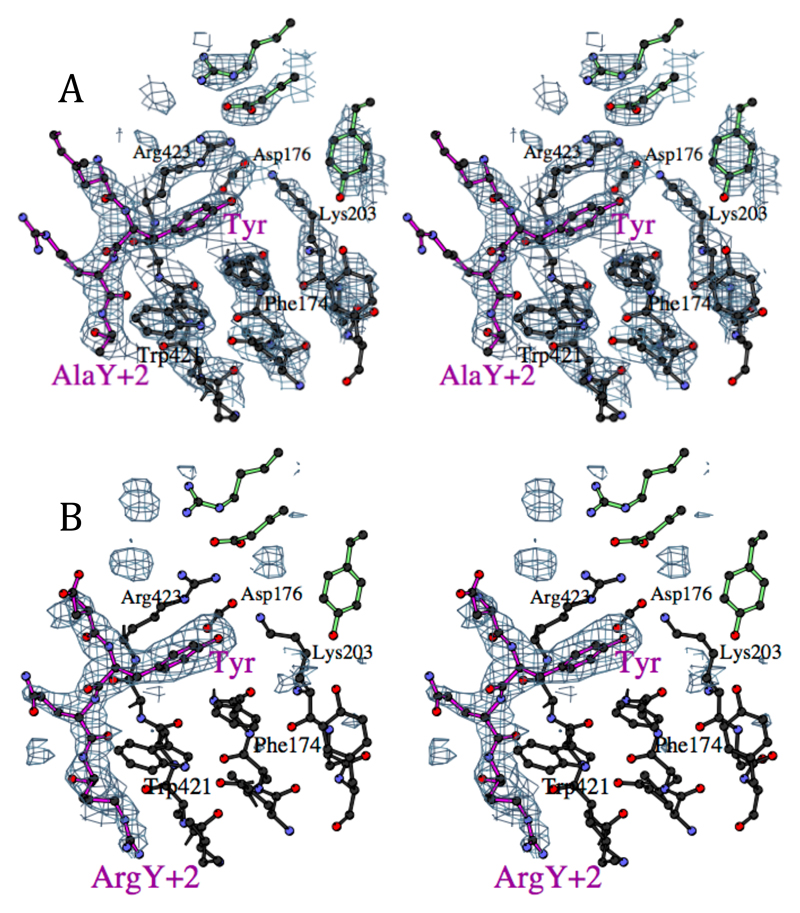
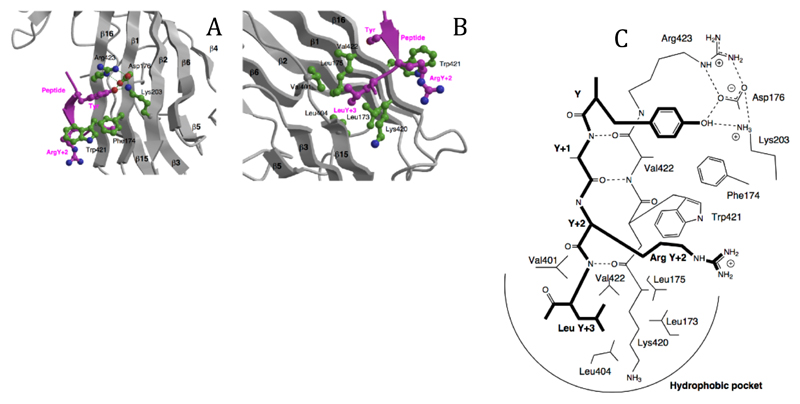
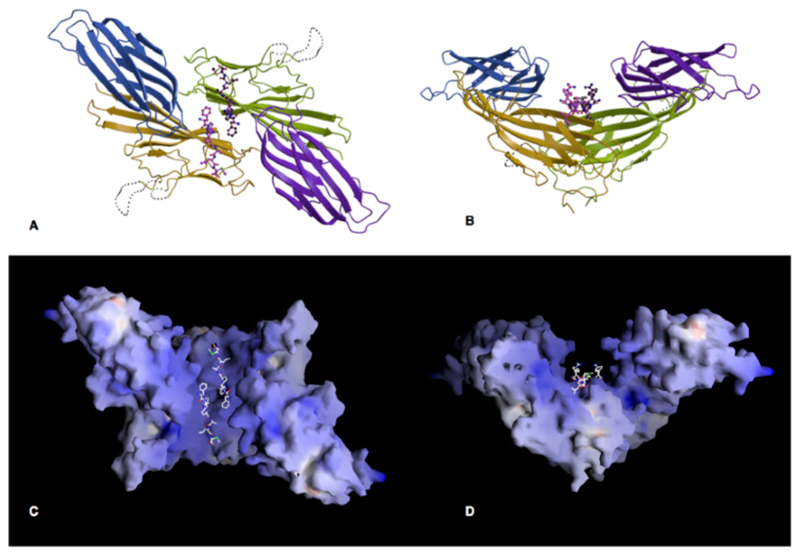
Many cell surface proteins are marked for endocytosis by a cytoplasmic sequence motif, tyrosine-X-X-(hydrophobic residue), that is recognized by the mu2 subunit of AP2 adaptors. Crystal structures of the internalization signal binding domain of mu2 complexed with the internalization signal peptides of epidermal growth factor receptor and the trans-Golgi network protein TGN38 have been determined at 2.7 angstrom resolution. The signal peptides adopted an extended conformation rather than the expected tight turn. Specificity was conferred by hydrophobic pockets that bind the tyrosine and leucine in the peptide. In the crystal, the protein forms dimers that could increase the strength and specificity of binding to dimeric receptors.


Figures

Figure 1
Figure 1. The structure
A,B Orthogonal views of μ2 with subdomain A shown in gold, subdomain B in blue and the peptide in magenta. Dotted lines represent disordered loops. The strands of the β-sheet (arrows) are numbered. The two subdomains are linked into a continuous β-sheet through strands 14 and 16/17. C Sequence alignment of μ2 from rat (Rat), human (Humn), Drosophila (Dros), C. elegans (cElg), Dictostylium (Dict), Arabidopsis thaliana (Plnt), S. pombe (Spmb), μ1 (AP47) from rat and μ3A (p47A) from rat. Identical residues are shaded red, conserved gold and those involved in internalisation signal binding in blue.
Figure 2
Figure 2
A Stereo view of the binding site for the tyrosine residue in the EGFR internalisation signal FYRALM, showing part of the experimental electron density map, with phases calculated using the peptide complex data as native with the Xe and EMTS derivatives, and solvent flattening with a 70% solvent content. The peptide is represented with magenta bonds, and the residues at the top right with green bonds come from the other subunit in the crystallographic dimer. (Figures drawn with BOBSCRIPT (32)) B Stereo view of the binding site for the TGN38 internalisation signal DYQRLN, in the same view as D. The difference electron density shown was calculated using the model from the FYRALM peptide structure with the peptide removed: density for the arginine in the Y+2 position is clearly visible, packed against Trp421.
Figure 3
Figure 3. The peptide binding site.
A The binding of the tyrosine residue of the internalisation signal peptide is in a hydrophobic pocket created by Phe174, Trp421 and Arg423, with a hydrogen-bonding network between the tyrosine OH and Asp176, Lys203 and Arg423. The structure shown is that of the DYQRLN TGN38 peptide. B The binding pocket for the bulky hydrophobic residue at Y+3 (Leucine in both peptides) is lined with aliphatic sidechains of Leu173, Leu175, Val401, Leu404, Val422 and the aliphatic portion of Lys420. ArgY+2 of the TGN38 peptide is packed against Trp421. C Schematic representation of the interactions between the internalisation signal of TGN38 and μ2, showing both side chain contacts and the short stretch of β-sheet formed between the peptide and β-strand 16. The peptide is shown with bold lines.
Figure 4
Figure 4. The crystallographic dimer
A, B Orthogonal views of the dimer formed in the crystal, along and perpendicular to the crystallographic twofold axis. The A subdomains are coloured gold and green and the B domains blue and purple. C and D. The surface of the μ2 dimer coloured according to electrostatic surface potential (blue positive, red negative, scale from -30 to +30 kT e-1)), in the same view as A and B. The planar face at the top of D may interact with the membrane. (Drawn with GRASP(33))

References

    1. Kirchausen T, Bonifacino JS, Riezman H. Curr Op Cell Biology. 1997;9:488. - PubMed
    1. Schlessinger J, Ullrich A. Neuron. 1992;9:383. - PubMed
    1. Chen WS, et al. Cell. 1989;59:33. - PubMed
    1. Wells A, et al. Science. 1990;247:962. - PubMed
    1. Schmid SL. Annu Rev Biochem. 1997;66:511. - PubMed

MeSH terms