. 1998 Nov 2;143(3):637-44.
doi: 10.1083/jcb.143.3.637.

Polyunsaturated fatty acids inhibit T cell signal transduction by modification of detergent-insoluble membrane domains

Affiliations

Polyunsaturated fatty acids inhibit T cell signal transduction by modification of detergent-insoluble membrane domains

T M Stulnig et al. J Cell Biol. .

Abstract

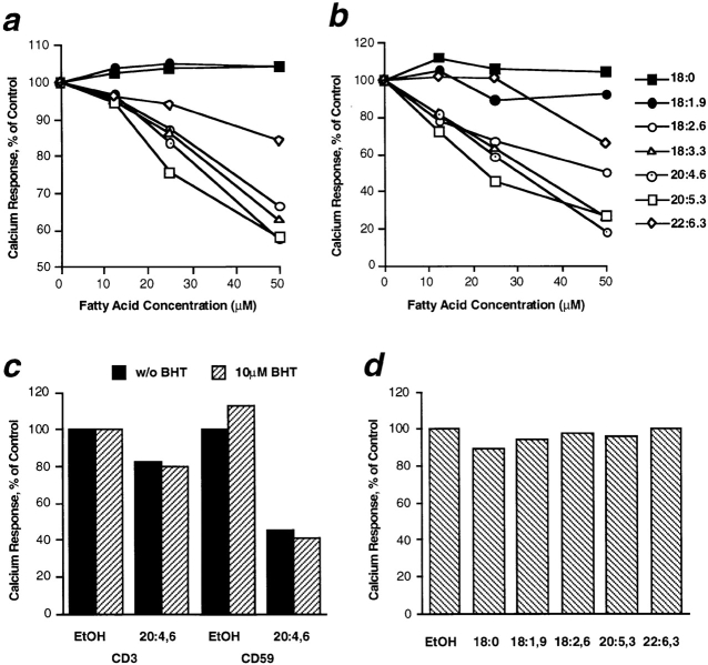
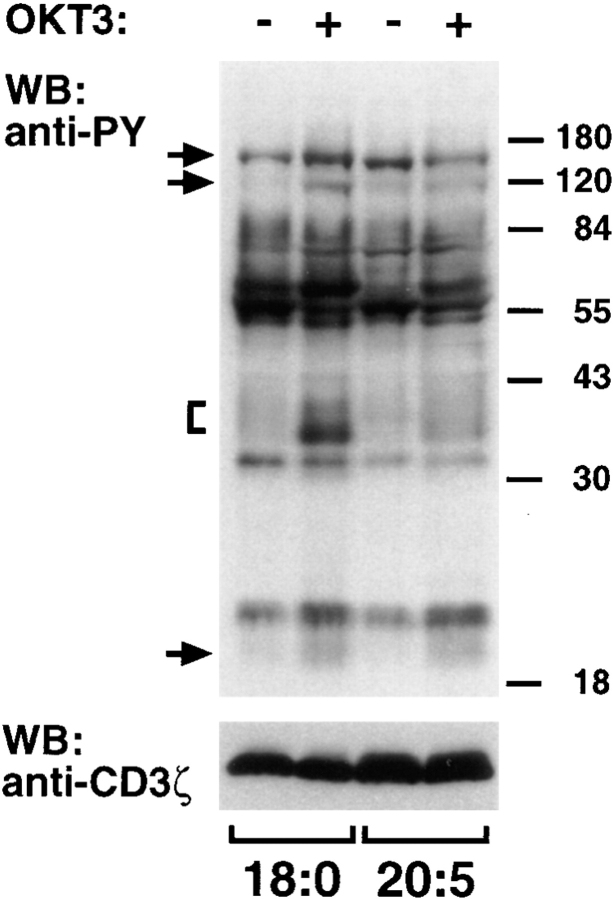
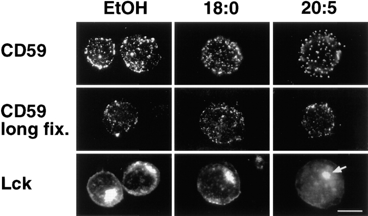
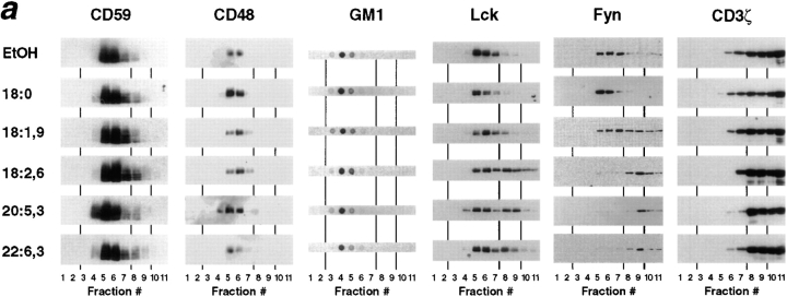
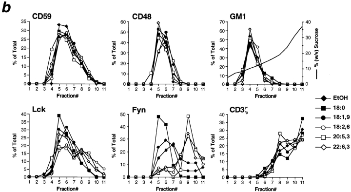
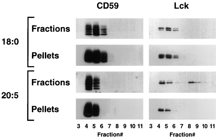
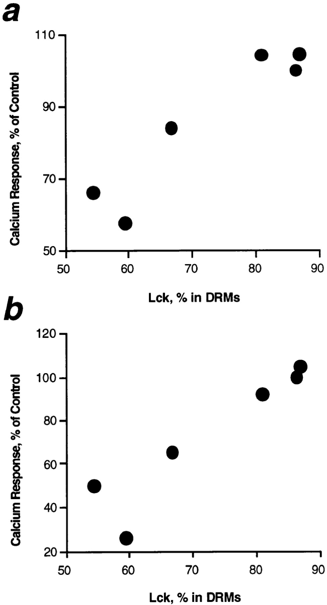
Polyunsaturated fatty acids (PUFAs) exert immunosuppressive effects, but the molecular alterations leading to T cell inhibition are not yet elucidated. Signal transduction seems to involve detergent-resistant membrane domains (DRMs) acting as functional rafts within the plasma membrane bilayer with Src family protein tyrosine kinases being attached to their cytoplasmic leaflet. Since DRMs include predominantly saturated fatty acyl moieties, we investigated whether PUFAs could affect T cell signaling by remodeling of DRMs. Jurkat T cells cultured in PUFA-supplemented medium showed a markedly diminished calcium response when stimulated via the transmembrane CD3 complex or glycosyl phosphatidylinositol (GPI)- anchored CD59. Immunofluorescence studies indicated that CD59 but not Src family protein tyrosine kinase Lck remained in a punctate pattern after PUFA enrichment. Analysis of DRMs revealed a marked displacement of Src family kinases (Lck, Fyn) from DRMs derived from PUFA-enriched T cells compared with controls, and the presence of Lck in DRMs strictly correlated with calcium signaling. In contrast, GPI-anchored proteins (CD59, CD48) and ganglioside GM1, both residing in the outer membrane leaflet, remained in the DRM fraction. In conclusion, PUFA enrichment selectively modifies the cytoplasmic layer of DRMs and this alteration could underlie the inhibition of T cell signal transduction by PUFAs.


Figures

Figure 1
Figure 1
Calcium signaling in T cells enriched with different fatty acids. Jurkat T cells were cultured in medium supplemented with distinct fatty acids as indicated, and stimulated for calcium response via CD3 or GPI-anchored CD59. The maximal rise in cytoplasmic calcium concentration was monitored and is given as percent of the solvent control (EtOH; Stulnig et al., 1997). (a and b) Cells incubated with various amounts of fatty acids were stimulated via CD3 (a) or CD59 (b). Means from three independent analyses are given for each fatty acid. (c) T cells were cultured with or without 25 μM polyunsaturated arachidonic acid (20:4 (n-6)) in presence or absence of the antioxidant BHT (10 μM) were stimulated via CD3 or CD59 as indicated. (d) T cells cultured with 25 μM of different fatty acids or EtOH were incubated with thapsigargin for induction of maximal calcium influx.
Figure 2
Figure 2
Stimulation of protein tyrosine phosphorylation in PUFA-enriched T cells. Jurkat T cells were cultured in medium supplemented with 50 μM eicosapentaenoic (20:5 (n-3)) or stearic acid (18:0) which served as control. Cells were either left unstimulated or stimulated by mAb OKT3 for 2 min as indicated. Lysates from 4 × 10⁵ cells per lane (21.6 and 21.2 μg cell protein for 18:0 and 20:5-treated cells, respectively) were analyzed by Western blotting for tyrosine phosphorylated proteins (PY), and for CD3ζ to control for equal loading. Note that the phosphorylation of typical phosphoproteins as indicated on the left was markedly abolished in stimulated PUFA-enriched T cells compared with the control. Molecular mass of marker proteins are given in thousands on the right.
Figure 3
Figure 3
Distribution of GPI-anchored CD59 and Src family kinase Lck in fatty acid–modified T cells. Jurkat T cells cultured in the presence of either no fatty acid (EtOH), 25 μM saturated stearic acid (18:0) or polyunsaturated eicosapentaenoic acid (20:5 (n-3)) were fixed with paraformaldehyde and analyzed for the cell surface distribution of CD59 by immunofluorescence. CD59 was detected on cells fixed by conventional formaldehyde fixation (CD59; mAb MEM-43), or by prolonged fixation that was proposed to prevent antibody-induced clustering of surface proteins (Mayor et al., 1994), but markedly interfered with immunodetection (CD59 long fix.; mAb MEM-43/5). For detection of Lck, fatty acid–modified cells were fixed in acetone before immunofluorescence staining. According to Ley et al. (1994), Lck was localized on the plasma membrane and in pericentrosomal vesicles (arrow). Compared with the clustered pattern of Lck in untreated (EtOH) and 18:0-treated cells, Lck was rather diffusely distributed in PUFA-enriched T cells but without discrimination of the nucleus suggesting membrane localization of the kinase. Bar, 5 μm.
Figure 4
Figure 4
Density gradient distribution of membrane constituents from fatty acid–modified T cells after lysis with nonionic detergent. (a) Membranes of T cells treated with 50 μM fatty acids were solubilized in lysis buffer containing 1% Brij-58 and fractionated by sucrose density gradient centrifugation. Floating fractions 4–7 indicate DRMs, fractions 8 and 9 were named intermediate, whereas most soluble proteins were localized at the bottom in fractions 10 and 11. Typical distribution of GPI-anchored proteins CD59 and CD48, ganglioside GM1, Src family kinases Lck and Fyn, and CD3ζ are shown from a single experiment from a total of 3–8 for each antigen. CD3ζ-blots were overexposed to illustrate its occurrence in DRM fractions. (b) Densitometric analysis of films shown in panel (a) with data from individual fractions given in percent of the whole gradient. The shape of the sucrose gradient is illustrated by the plain line within the GM1 histogram.
Figure 4
Figure 4
Density gradient distribution of membrane constituents from fatty acid–modified T cells after lysis with nonionic detergent. (a) Membranes of T cells treated with 50 μM fatty acids were solubilized in lysis buffer containing 1% Brij-58 and fractionated by sucrose density gradient centrifugation. Floating fractions 4–7 indicate DRMs, fractions 8 and 9 were named intermediate, whereas most soluble proteins were localized at the bottom in fractions 10 and 11. Typical distribution of GPI-anchored proteins CD59 and CD48, ganglioside GM1, Src family kinases Lck and Fyn, and CD3ζ are shown from a single experiment from a total of 3–8 for each antigen. CD3ζ-blots were overexposed to illustrate its occurrence in DRM fractions. (b) Densitometric analysis of films shown in panel (a) with data from individual fractions given in percent of the whole gradient. The shape of the sucrose gradient is illustrated by the plain line within the GM1 histogram.
Figure 5
Figure 5
Detergent-insolubility of gradient fractions in fatty acid– treated T cells. Membranes of Jurkat T cells treated with 50 μM stearic acid (18:0) or polyunsaturated eicosapentaenoic acid (20:5 (n-3)) were fractionated by density gradient centrifugation as described in Fig. 4. Aliquots of individual fractions were diluted and pelleted to test for detergent-insoluble components. CD59 and Lck were detected in fractions and pellets by Western blotting.
Figure 6
Figure 6
Correlation of T cell calcium response with the presence of Lck in DRMs. T cells were treated with 50 μM of individual fatty acids or ethanol alone as given in Fig. 4. The proportion of Src family kinase Lck recovered in DRM fractions 4–7 as shown in Fig. 4 is given on the abscissa, and means of calcium responses via CD3 (a) or GPI-anchored CD59 (b) are displayed on the ordinate. Note the close correlation between both parameters in all fatty acid treatments.

References

    1. Ahmed SN, Brown DA, London E. On the origin of sphingolipid/ cholesterol rich detergent insoluble cell membranes: physiological concentrations of cholesterol and sphingolipid induce formation of a detergent insoluble, liquid ordered lipid phase in model membranes. Biochemistry. 1997;36:10944–10953. - PubMed
    1. Arni S, Ilangumaran S, van Echten G, Deckert, Sandhoff K, Poincelet M, Briol A, Rungger E, Brandle, Hoessli DC. Differential regulation of Src-family protein tyrosine kinases in GPI domains of T lymphocyte plasma membranes. Biochem Biophys Res Commun. 1996;225:801–807. - PubMed
    1. Belluzzi A, Brignola C, Campieri M, Pera A, Boschi S, Miglioli M. Effect of an enteric-coated fish-oil preparation on relapses in Crohn's disease. N Engl J Med. 1996;334:1557–1560. - PubMed
    1. Bligh EG, Dyer WJ. A rapid method for total lipid extraction and purification. Can J Biochem Physiol. 1959;37:911–917. - PubMed
    1. Bohuslav J, Cinek T, Hoř ejší V. Large, detergent-resistant complexes containing murine antigens Thy-1 and Ly-6 and protein tyrosine kinase p56lck. Eur J Immunol. 1993;23:825–831. - PubMed

Publication types

Substances