. 1998 Nov 2;143(3):673-85.
doi: 10.1083/jcb.143.3.673.

Localization of the kinesin-like protein Xklp2 to spindle poles requires a leucine zipper, a microtubule-associated protein, and dynein

Affiliations

Localization of the kinesin-like protein Xklp2 to spindle poles requires a leucine zipper, a microtubule-associated protein, and dynein

T Wittmann et al. J Cell Biol. .

Abstract

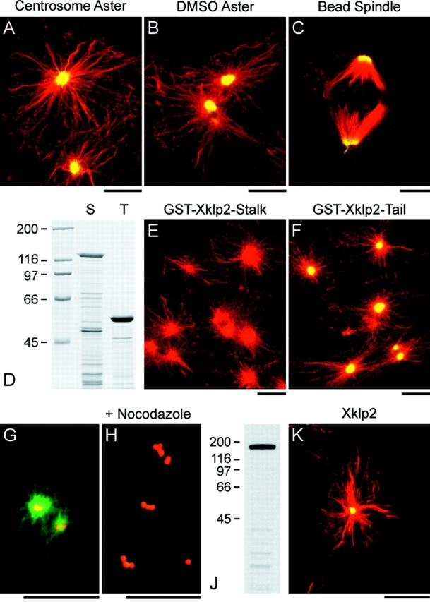
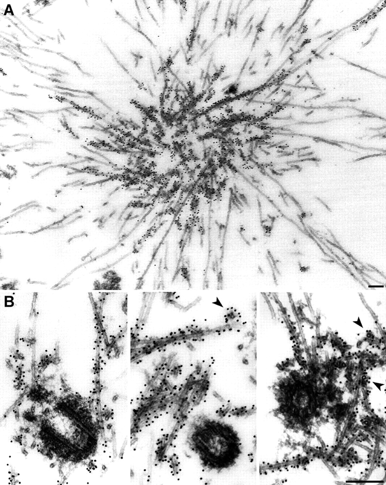
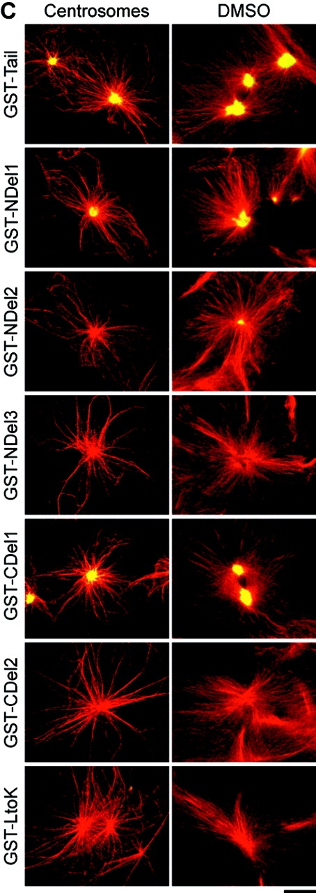
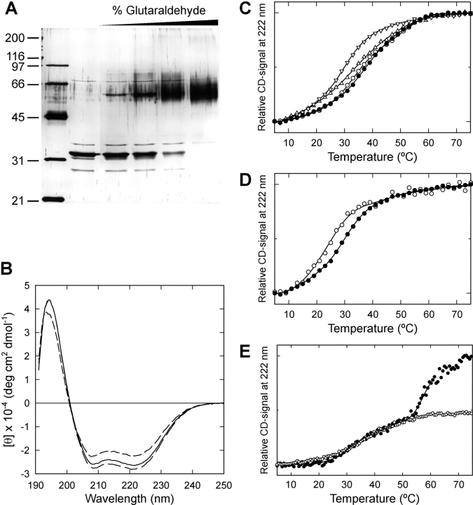
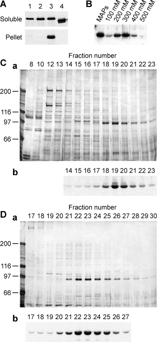
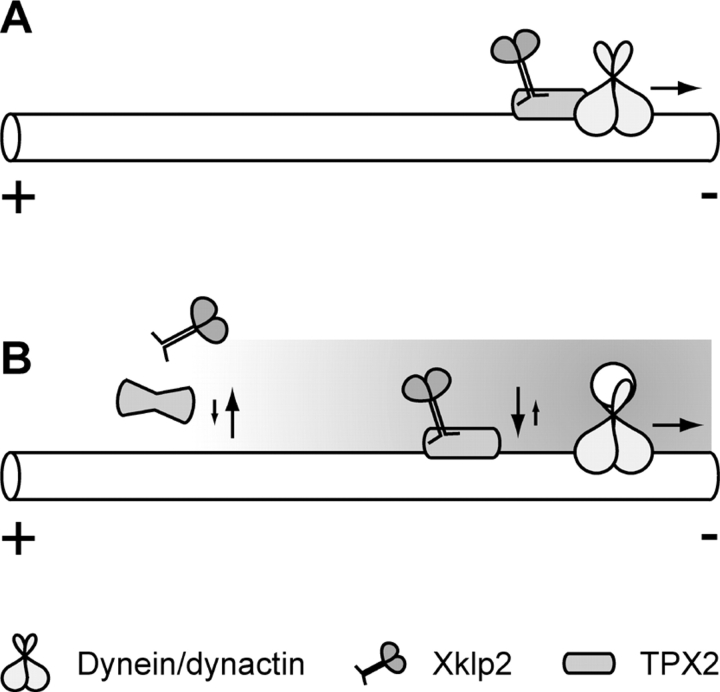
Xklp2 is a plus end-directed Xenopus kinesin-like protein localized at spindle poles and required for centrosome separation during spindle assembly in Xenopus egg extracts. A glutathione-S-transferase fusion protein containing the COOH-terminal domain of Xklp2 (GST-Xklp2-Tail) was previously found to localize to spindle poles (Boleti, H., E. Karsenti, and I. Vernos. 1996. Cell. 84:49-59). Now, we have examined the mechanism of localization of GST-Xklp2-Tail. Immunofluorescence and electron microscopy showed that Xklp2 and GST-Xklp2-Tail localize specifically to the minus ends of spindle pole and aster microtubules in mitotic, but not in interphase, Xenopus egg extracts. We found that dimerization and a COOH-terminal leucine zipper are required for this localization: a single point mutation in the leucine zipper prevented targeting. The mechanism of localization is complex and two additional factors in mitotic egg extracts are required for the targeting of GST-Xklp2-Tail to microtubule minus ends: (a) a novel 100-kD microtubule-associated protein that we named TPX2 (Targeting protein for Xklp2) that mediates the binding of GST-Xklp2-Tail to microtubules and (b) the dynein-dynactin complex that is required for the accumulation of GST-Xklp2-Tail at microtubule minus ends. We propose two molecular mechanisms that could account for the localization of Xklp2 to microtubule minus ends.


Figures

Figure 1
Figure 1
Localization of GST-Xklp2-Tail is cell cycle dependent. (A) Asters were assembled around human centrosomes in 10,000 g mitotic egg extract (a–c), the same extract released into interphase by the addition of calcium and cycloheximide (d–f), and cycled back to mitosis by addition of cyclin Δ90 (g–i). The asters were assembled in the presence of rhodaminated tubulin (a, d, and g), and stained with a human anti-centrosome antibody (b, e, and h), and an anti-GST antibody (c, f, and i) that were revealed with FITC- and Cy5-conjugated secondary antibodies, respectively. GST-Xklp2-Tail accumulates at the center of mitotic asters (a–c). This localization is abolished during interphase (d–f) and reappears after reentry into mitosis (g–i). (B) Asters assembled in the presence of 5% DMSO in 150,000 g mitotic egg extract (a and b), and microtubules in the same extract released to interphase before addition of DMSO (c and d). (a and c) Microtubules, (b and d) anti-GST-staining. GST-Xklp2-Tail is strongly concentrated at the center of mitotic DMSO asters (b) and stained diffusely along microtubules in interphase (d). All reactions contained 2 μM GST-Xklp2-Tail. Bars, 10 μm.
Figure 2
Figure 2
Both endogenous Xklp2 and GST-Xklp2-Tail localize to microtubule minus ends in mitotic asters in the absence of centrosomes. (A–C) Localization of GST-Xklp2-Tail. Microtubules are red, anti-GST-staining is green. (A) Aster nucleated by human centrosomes, (B) Aster assembled in the presence of 5% DMSO, (C) Spindle assembled around chromatin beads (Heald et al., 1996). Although there are no centrosomes present in B and C, GST-Xklp2-Tail accumulates at the center of the asters or the spindle poles, respectively. (D–F) The COOH-terminal domain of Xklp2 is necessary and sufficient for localization. (D) Coomassie-stained SDS-PAGE of GST-Xklp2-Stalk (amino acids 363–1137, S) and GST-Xklp2-Tail (amino acids 1137–1387, T). GST-Xklp2-Stalk does not localize to the center of taxol-induced mitotic asters (E) whereas GST-Xklp2-Tail does in a parallel experiment (F). (G and H) Depolymerization of microtubules eliminates GST-Xklp2-Tail staining around centrosomes. Centrosomes are red, anti-GST-staining is green, microtubules are not shown. (G) Aster nucleated by human centrosomes in the presence of 2 μM GST-Xklp2-Tail and stained with the human anti-centrosome antibody, (H) Asters assembled as in G, but treated with 5 μM nocodazole and incubated for 5 min on ice before fixation. (J) Western blot of 0.5 μl egg extract probed with an affinity-purified anti-Xklp2-Tail antibody. The antibody strongly recognizes a band of 160 kD. (K) Localization of the endogenous Xklp2. Microtubules are red, anti-Xklp2-staining is green. Asters were assembled with 5% DMSO in the presence of FITC-labeled tubulin and stained with the anti-Xklp2-Tail antibody (∼5 μg/ml) and a Cy3-conjugated secondary antibody. The anti-Xklp2-Tail antibody stained the center of the DMSO-asters. Bars, 10 μm.
Figure 3
Figure 3
Localization of GST-Xklp2-Tail by immunoelectron microscopy. (A) Aster nucleated by purified human centrosomes in mitotic 10,000 g egg extract in the presence of 2 μM GST-Xklp2-Tail, stained with an anti-GST-antibody. The centrosomes are not visible in this section. (B) Centrosomes in the center of mitotic asters from the same sample. Gold particles were observed along microtubule minus ends but not on centrosomes. Arrowheads point to microtubules in cross-section, revealing the distance between gold particles and the microtubules. We did not observe any labeling in negative controls when the primary antibody was omitted or when the mutant fusion protein missing the leucine zipper (GST-Xklp2-CDel2) was added to the reaction instead of the tail (data not shown). Bars, 200 nm.
Figure 4
Figure 4
Biochemical analysis of Xklp2 cosedimentation with microtubules. Taxol (1 μM) and the m70.1 antibody (1–2 mg/ml) were added as indicated to 150,000 g egg extracts and the reactions incubated for 30 min at 20°C. M, mitotic extract, I, interphase extract. Microtubule pellets (corresponding to 5 μl of extract) and soluble fractions (corresponding to 0.25 μl of extract) were analyzed on Coomassie-stained gels and Western blots probed for the proteins indicated (DIC, dynein intermediate chain). (A) Sedimentation of endogenous Xklp2. (B) Sedimentation when either GST-Xklp2-Tail, T, or GST-Xklp2-LtoK, K, were added to the extract at 0.3 μM. Xklp2 as well as the GST-Xklp2-Tail sediment with microtubules in a mitotic cytoplasm. GST-Xklp2-Tail (52 kD) runs very close to the tubulin doublet. This is why the band on the Western blot of the microtubule pellet appears broadened. Also the dynein intermediate chain is enriched in mitotic microtubule pellets. When the m70.1 antibody is added the dynein intermediate chain is not associated with microtubules anymore and the binding of Xklp2 and GST-Xklp2-Tail to microtubules is reduced. The molecular mass of marker proteins is indicated on the right.
Figure 5
Figure 5
Localization of the truncated GST-Xklp2-Tail fusion proteins to mitotic asters. (A) Coiled-coil prediction for the Xklp2-Tail obtained with the Coils- (solid line; Lupas et al., 1991) and the Paircoil-algorithm (dashed line; Berger et al., 1995), and schematic representation of the different constructs. Vertical bars represent the four leucines present in the leucine zipper. (B) Coomassie Brilliant blue–stained SDS-gel of the purified GST-fusion proteins and the thrombin-cleaved proteins used for circular dichroism spectroscopy. The molecular mass of marker proteins is indicated on the left. (C) Localization of the GST-fusion proteins to asters nucleated by human centrosomes in 10,000 g egg extract and to DMSO-asters in 150,000 g egg extract. The fusion proteins were added at a concentration of 2 μM and are indicated on the left. Both the NH2-terminal region and the COOH-terminal leucine zipper are required for localization of the GST-Xklp2-Tail to the center of mitotic asters. Microtubules are red, anti-GST-staining is green. Bar, 10 μm.
Figure 5
Figure 5
Localization of the truncated GST-Xklp2-Tail fusion proteins to mitotic asters. (A) Coiled-coil prediction for the Xklp2-Tail obtained with the Coils- (solid line; Lupas et al., 1991) and the Paircoil-algorithm (dashed line; Berger et al., 1995), and schematic representation of the different constructs. Vertical bars represent the four leucines present in the leucine zipper. (B) Coomassie Brilliant blue–stained SDS-gel of the purified GST-fusion proteins and the thrombin-cleaved proteins used for circular dichroism spectroscopy. The molecular mass of marker proteins is indicated on the left. (C) Localization of the GST-fusion proteins to asters nucleated by human centrosomes in 10,000 g egg extract and to DMSO-asters in 150,000 g egg extract. The fusion proteins were added at a concentration of 2 μM and are indicated on the left. Both the NH2-terminal region and the COOH-terminal leucine zipper are required for localization of the GST-Xklp2-Tail to the center of mitotic asters. Microtubules are red, anti-GST-staining is green. Bar, 10 μm.
Figure 6
Figure 6
Dimerization of the Xklp2-Tail. (A) Silver-stained SDS-PAGE of thrombin-cleaved Xklp2-Tail incubated with increasing amounts of glutaraldehyde (from left to right: markers, 0, 0.0003, 0.001, 0.003, and 0.01%). With rising glutaraldehyde concentrations, a new band appears at 60 kD corresponding to the dimeric Xklp2-Tail. The molecular mass of marker proteins is indicated on the left. (B) Far UV circular dichroism spectra of Xklp2-Tail (solid line), CDel2 (long dash), and NDel2 (short dash) at 2 μM concentration at 4°C. (C) Thermal unfolding of Xklp2-Tail (filled circle), CDel2 (open circle), NDel1 (triangle), NDel2 (upsidedown triangle) at 10 μM. (D) Thermal unfolding of Xklp2-NDel2 at a concentration of 10 μM (filled circle) and 1 μM (open circle). (E) Thermal unfolding of GST-Xklp2-Tail (filled circle) and Xklp2-Tail (open circle) at 10 μM concentration. See text for details.
Figure 7
Figure 7
Cytoplasmic dynein and the dynactin complex are required for localization of GST-Xklp2-Tail. (A) Human centrosomes were added to 10,000 g mitotic egg extract and incubated at 20°C. After 10 min, 1–2 mg/ml m70.1 antibody was added to b, after another 5 min 2 μM GST-Xklp2-Tail was added and the reaction incubated for 45 min. (d) 1–2 mg/ml m70.1 was added to 150,000 g egg extract and incubated for 5 min on ice. Microtubule polymerization was then induced by addition of 5% DMSO and incubation for 30 min at 20°C. In both cases addition of m70.1 disrupts the localization of GST-Xklp2-Tail to the center of mitotic asters. (B) Disruption of the dynactin complex inhibits localization of GST-Xklp2-Tail. (b) Asters nucleated by human centrosomes in the presence of partially purified bacterially expressed p50/dynamitin (∼1 mg/ml) in 10,000 g mitotic egg extract for 60 min at 20°C. (d) Asters assembled by 1 μM taxol in the presence of 1 mg/ml p50/dynamitin in 10,000 g mitotic egg extract for 30 min at 20°C. Microtubules are red, anti-GST-staining is green. Bars, 10 μm.
Figure 8
Figure 8
Purification of TPX2, a MAP that mediates the binding of Xklp2 to microtubules. (A) GST-Xklp2-Tail alone does not bind to pure taxol-stabilized microtubules (lane 1). However, a fraction of MAPs contains an activity that mediates the binding of GST-Xklp2-Tail to microtubules (lane 3). This activity was assayed in the following way: lane 1, prepolymerized microtubules were mixed with either GST-Xklp2-Tail and control buffer; lane 2, GST-Xklp2-Tail and soluble proteins (0.6 mg/ml) from CSF-arrested egg extract; lane 3, GST-Xklp2-Tail and MAPs (0.6 mg/ ml); lane 4, or GST-Xklp2-CDel2 and MAPs. The microtubules were sedimented by centrifugation through a sucrose cushion and the soluble fractions and pellets (four times more loaded than of the soluble fraction) were analyzed by Western blotting, probed with an anti-GST antibody. (B) Fractionation of MAPs by sequential salt elution from microtubules. Microtubules polymerized in CSF-arrested egg extract were purified by centrifugation through a sucrose cushion and eluted in one step with 500 mM NaCl (MAPs) or sequentially with the NaCl concentration indicated. The fractions were assayed as described above and the pellets were analyzed by Western blotting and probed with an anti-GST antibody. (C) Mono S chromatography of the 300 mM NaCl fraction eluted from microtubules. (a) Silver-stained SDS-PAGE of 5-μl aliquots of the fractions indicated. The molecular mass of marker proteins is indicated on the left. (b) Assay for the activity mediating the binding of GST-Xklp2-Tail to microtubules of fractions from the Mono S chromatography as indicated. (D) The peak fraction of the Mono S chromatography was applied to a Superdex 200 gel filtration column. (a) Silver-stained gel of 5-μl aliquots of the fractions indicated. (b) Assay for the activity mediating the binding of GST-Xklp2-Tail to microtubules of fractions from the Superdex 200 gel filtration chromatography as indicated. In both chromatographic steps the activity copurified with a 100-kD protein.
Figure 9
Figure 9
Two alternative models for how Xklp2 and TPX2 could be localized to microtubule minus ends in a dynein dependent way. (A) Direct mechanism: Xklp2 and TPX2 interact with a dynein containing complex that moves towards the microtubule minus end or they bind directly to a component that has been transported there by dynein before. (B) Indirect mechanism: a protein that is transported to the minus ends by dynein creates a gradient along the microtubule (possibly by some enzymatic activity) that changes the affinity of TPX2 to Xklp2 and/or to microtubules.

References

    1. Afshar K, Barton NR, Hawley RS, Goldstein LS. DNA binding and meiotic chromosomal localization of the Drosophilanod kinesin-like protein. Cell. 1995a;81:129–138. - PubMed
    1. Afshar K, Scholey J, Hawley RS. Identification of the chromosome localization domain of the DrosophilaNod kinesin-like protein. J Cell Biol. 1995b;131:833–843. - PMC - PubMed
    1. Andersen SSL, Karsenti E. XMAP310: A Xenopusrescue-promoting factor localized to the mitotic spindle. J Cell Biol. 1997;139:975–983. - PMC - PubMed
    1. Ashford, A.J., S.S.L. Andersen, and A.A. Hyman. 1998. Preparation of tubulin from bovine brain. In Cell Biology: A Laboratory Handbook. Vol. 2. J.E. Celis, editor. Academic Press, San Diego, CA. 205–212.
    1. Barton NR, Goldstein LSB. Going mobile: microtubule motors and chromosome segregation. Proc Natl Acad Sci USA. 1996;93:1735–1742. - PMC - PubMed